Digital workflows are increasingly a part of today’s dental practices and dental laboratories (1). Digital technology offers clinical and technical advantages that traditional methods could not guarantee in the past (2).
For example, the ability to digitally superimpose two models of the dental arches of the same patient allows us to observe differences, changes and wear patterns that were once only assessable visually and in a completely arbitrary and subjective way.
This has allowed not only more precise comparisons, but also the development of new procedures and workflows, such as virtual cross mounting, a technique useful for maintaining the same intermaxillary relationship when moving from a removable total denture to a fixed one on implants (3,4).
These virtual overlays also allow you to visualise tooth movements during orthodontic treatments, verifying the correspondence between the clinical evolution and the virtual setup (5).
Where analogue technology remains irreplaceable in dentistry
However, there are still areas where analogue is difficult to replace with digital due to technological and functional limitations (6).
This is true, for example, of removable prostheses, where – as discussed in our previous articles (7,8) – impressions are currently more reliable if taken with analogue techniques and materials with different viscosities and consistencies. In fact, these adapt better to the soft tissues in cases of complete anodontia by correctly stretching the mucosa on the underlying bone bases (9). Furthermore, recording the intermaxillary relationship in these patients is not yet achievable solely through intraoral scanners.
Several techniques have been proposed to overcome these difficulties in cases of implant-supported prostheses. For example, using some references on the palate or the palate itself, two successive scans can be matched; one with the temporary prosthesis in situ and another with the scan bodies (10). This allows the technician to create a prosthesis on implants with the same occlusion as the previous temporary prosthesis.
Other techniques involve the use of scan bodies equipped with special pillars which can be adjusted to different heights and therefore allow the arches to be matched during the phase of digital bite registration (11).
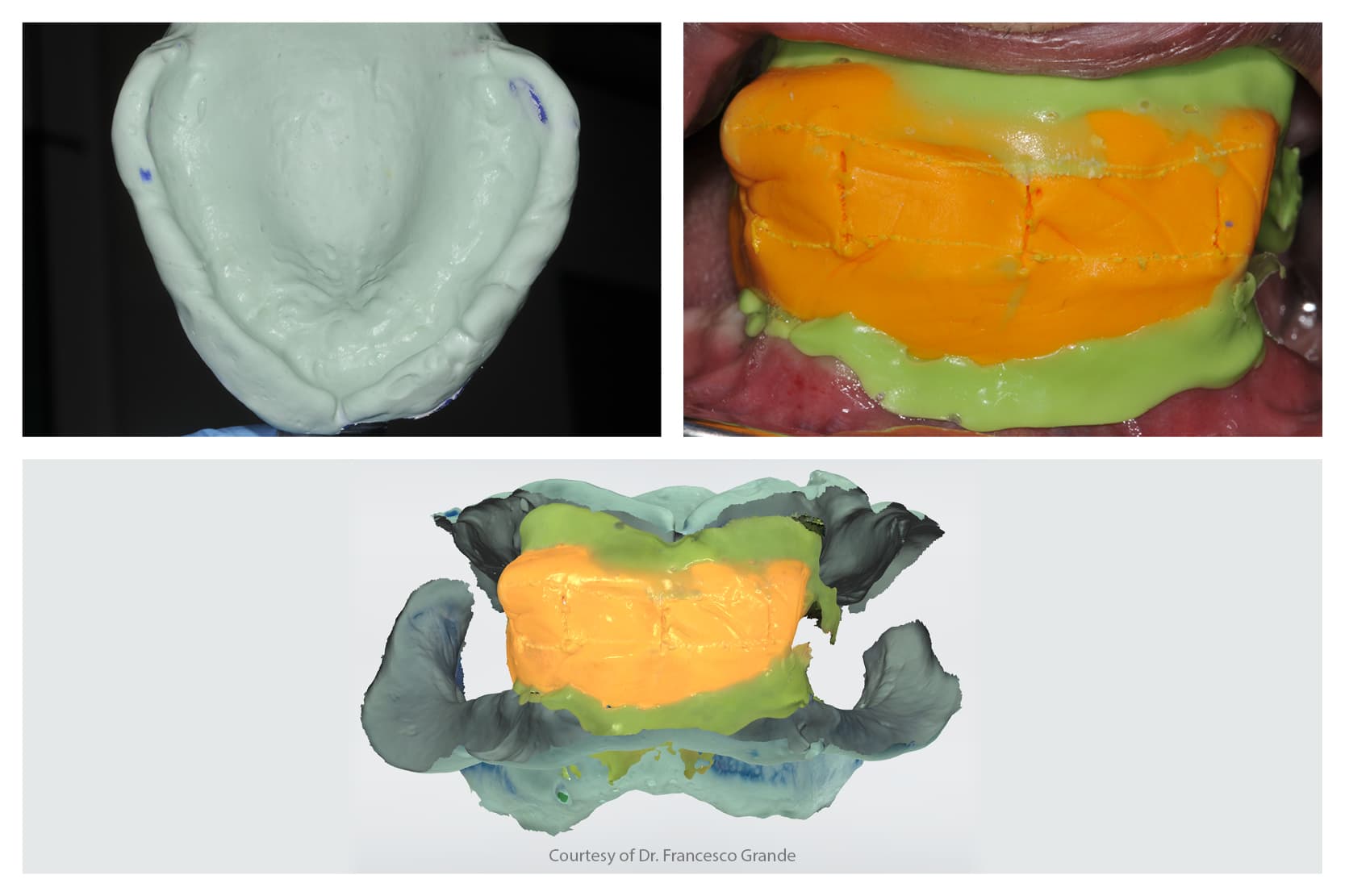
Regarding the articulation of totally edentulous jaws in removable prostheses, the scanning of a block of silicone putty placed between the arches at the desired vertical dimension has been proposed (12,13). This block can then be scanned and matched to intraoral scans or conventional impression scans (12,13).
It can also be used to articulate the plaster models of the respective arches, thereby allowing the technician to start working at an initial vertical dimension.
The putty block can also be characterised with reference lines – such as the midline, smile line and canine line – to guide the positioning of the anterior teeth, which will then be reviewed or confirmed clinically if necessary (13).
Combination of analogue and digital techniques in dentistry
Another step in which the combination of analogue and digital technologies proves successful is provided by the Wagner Try-Ins. These are represented by 3D printed bases and wax teeth at the base. Unlike traditional wax ridges, they are not made on physical plaster models, appropriately relieved in the undercut areas to avoid being damaged by the continuous insertion and removal of the artefacts, but are produced entirely digitally.
Since they can also exploit any undercuts, they are more stable in the oral cavity. Furthermore, with the waxed teeth on the printed base, they offer the option of freely modifying their position and evaluating their aesthetics more effectively than prototypes printed in a single block with a uniform white colour (14).
As regards the production of removable prostheses, it must be noted that digital techniques and in particular milling are beneficial as they are not affected by contraction due to polymerisation, porosity and monomeric loss typical of removable prostheses made using analogue techniques (15,16).
However, one problem can be the aesthetics of milled or printed teeth which are not as aesthetic as splint teeth due to the different vestibular-lingual translucencies present in the latter. A hybrid solution combining a milled base with preformed splint teeth has recently been proposed in a study and could provide an alternative while waiting for the materials used for printing or milling to be improved from an aesthetic point of view (13).
However, even with this solution there are disadvantages; in particular, the risk of individual teeth detaching from the socket where they are glued.
Analogue-digital integration in dental practices: the most effective approach
In conclusion, we can say that the integration between analogue and digital is the most effective strategy today for obtaining predictable, high-quality clinical results, especially in complex fields such as removable complete dentures.
Although digital technology offers tools to simplify, document and improve workflows, some phases are still dominated by traditional techniques for reasons of reliability and adaptability to clinical situations.
References:
Images extracted from “CAD-CAM complete digital dentures: An improved clinical and laboratory workflow” – https://doi.org/10.1016/j.prosdent.2024.11.016
1. Revilla-Leon M, Frazier K, da Costa JB, Kumar P, Duong ML, Khajotia S, et al. Intraoral scanners: An American Dental Association Clinical Evaluators Panel survey. J Am Dent Assoc. 2021 Aug 1;152(8):669-670.e2.
2. Robles-Medina M, Romeo-Rubio M, Salido MP, Pradíes G. Digital Intraoral Impression Methods: an Update on Accuracy. Curr Oral Health Rep. 2020 Dec;7(4):361–75.
3. Lepidi L, Galli M, Grammatica A, Joda T, Wang HL, Li J. Indirect Digital Workflow for Virtual Cross-Mounting of Fixed Implant-Supported Prostheses to Create a 3D Virtual Patient. J Prosthodont. 2021 Feb;30(2):177–82.
4. Tallarico M, Galiffi D, Scrascia R, Gualandri M, Zadrożny Ł, Czajkowska M, et al. Digital Workflow for Prosthetically Driven Implants Placement and Digital Cross Mounting: A Retrospective Case Series. Prosthesis. 2022 Sep;4(3):353–68.
5. de Leotard A, Le Norcy E. Comparison of dental movements in digital setups created by orthodontists and ‘3shape design service®’ engineers: A cross-sectional study. Int Orthod. 2024 Dec;22(4):100919.
6. Revilla-León M, Lanis A, Yilmaz B, Kois JC, Gallucci GO. Intraoral digital implant scans: Parameters to improve accuracy. J Prosthodont. 2023 Dec;32(S2):150–64.
7. zhermack. Impronte Dentali Analogiche e Digitali a Confronto [Internet]. [cited 2025 Jun 21]. Available from: https://magazine.zhermack.com/it/studio/impronta-digitale-vs-impronta-analogica-quando-usare-luna-e-quando-laltra/
8. zhermack. Impronta analogica e digitale su paziente edentulo: review della letteratura [Internet]. [cited 2025 Jun 21]. Available from: https://magazine.zhermack.com/it/studio/impronta-analogica-vs-digitale-su-paziente-edentulo/
9. Chebib N, Kalberer N, Srinivasan M, Maniewicz S, Perneger T, Müller F. Edentulous jaw impression techniques: An in vivo comparison of trueness. J Prosthet Dent. 2019 Apr;121(4):623–30.
10. Chochlidakis K, Romeo D, Ercoli C, Papaspyridakos P. Complete Digital Workflow for Prosthesis Prototype Fabrication with the Double Digital Scanning (DDS) Technique: A Prospective Study on 16 Edentulous Maxillae. Journal of Prosthodontics [Internet]. 2022 [cited 2025 Apr 19];31(9):761–5. Available from: https://onlinelibrary.wiley.com/doi/abs/10.1111/jopr.13569
11. Nuytens P, Grande F, Li J, Lepidi L. Maxillomandibular relationship and virtual facebow integration in complete-arch intraoral implant scan: A novel clinical technique. J Prosthodont. 2024 Mar 20;
12. Srinivasan M, Kalberer N, Naharro M, Marchand L, Lee H, Müller F. CAD-CAM milled dentures: The Geneva protocols for digital dentures. J Prosthet Dent. 2020 Jan;123(1):27–37.
13. Grande F, Pavone L, Molinelli F, Mussano F, Srinivasan M, Catapano S. CAD-CAM complete digital dentures: An improved clinical and laboratory workflow. J Prosthet Dent. 2024 Dec 28;S0022-3913(24)00821-7.
14. Wagner SA, Kreyer R. Digitally Fabricated Removable Complete Denture Clinical Workflows using Additive Manufacturing Techniques. J Prosthodont. 2021 May;30(S2):133–8.
15. AlHelal A, AlRumaih HS, Kattadiyil MT, Baba NZ, Goodacre CJ. Comparison of retention between maxillary milled and conventional denture bases: A clinical study. J Prosthet Dent. 2017 Feb;117(2):233–8.
16. Grande F, Tesini F, Pozzan MC, Zamperoli EM, Carossa M, Catapano S. Comparison of the Accuracy between Denture Bases Produced by Subtractive and Additive Manufacturing Methods: A Pilot Study. Prosthesis. 2022 Mar 28;4(2):151–9.
Would you like more information about Zhermack Dental products and solutions?
Contact us


