The reproduction of soft tissues in implant-supported prostheses is a fundamental aspect in implant-prosthetic therapy as it contributes to achieving a harmonious and biomimetic aesthetic result through defining the emergence profile and interdental papillae (1).
At the same time, the peri-implant soft tissues are an indispensable biological barrier for protecting the supporting bone and preventing inflammatory complications, thereby ensuring functional stability and longevity of the implant-prosthetic restoration (1,2).
Implant supracrestal complex
For these reasons, in fact, some authors use the term implant supracrestal complex (ISC), which defines the anatomical and functional complex made up of the peri-implant soft tissues, the mechanical components and the bacterial flora that interact along the transmucosal tract of the implant-supported prosthesis (1).
This approach emphasises how the long-term success of implant-supported rehabilitations does not depend solely on osseointegration, but also on the dynamic balance that is established between the above-mentioned elements in maintaining health, aesthetics and functional stability (1,2).
Techniques for reproducing peri-implant soft tissues
Currently, the techniques for reproducing peri-implant soft tissues can be divided into two main categories: analogue and digital (3).
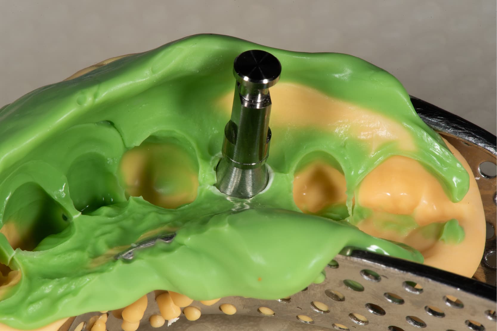
One of the best known analogue techniques allows us to copy the peri-implant soft tissue based on the shape of the temporary prosthesis (4). In this technique, we start with a temporary prosthesis that has already been adapted and has the correct emergence profiles.
After connecting the temporary prosthesis extraorally to the implant analogues, the emergence profile is taken using silicone putty. Subsequently, once the temporary prosthesis has been removed, the standard pick-up transfers are screwed onto the same analogues. The space that remains between the transfers and the internal walls of the silicone is then filled with resin, thereby customising the transfers. These will then be used for the final intraoral impression.
Customised transfers can also be made directly in the mouth (5) immediately after removing the temporary prosthesis, to prevent the tissues from being remodelled and losing the shape that has been created. In this technique, composite resin is injected around the copings and at the level of the intermediate elements, thereby accurately recreating the profile of the tissues and locking everything in the position achieved with the temporary prosthesis.
On the other hand, Londono et al. (6) use the temporary prosthesis itself as an impression transfer to accurately reproduce on the model the profiles of the tissues modelled in the mouth. To ensure it is correctly repositioned in the impression, a small blob of composite is applied to its vestibular surface, which acts as a marker allowing it to be inserted into the exact position during casting of the model.
If the patient does not have a temporary prosthesis before the final impression and the correct emergence profile needs to be transferred to the master model in a single session, Yilmaz offers an alternative analogue approach.
In this technique the temporary prosthesis, produced immediately after impression, is screwed onto the implant analogue of the model and used to shape the artificial gum directly, so that the model accurately reproduces the emergence profile created in the mouth (7).
However, according to the technique suggested by Lin et al. (8), a milled polyurethane model, based on an intraoral scan, is subsequently modified in an analogue way to reproduce the soft tissue profiles generated by the temporary prosthesis.
After taking a simple alginate impression with the temporary prosthesis in place, it is screwed onto the milled model and silicone is injected around it to obtain a removable replica of the peri-implant tissues.
This technique allows Lin to transfer the actual morphology of the soft tissues onto the digital impression model, integrating the benefits of CAD/CAM technology with the precision of the analogue technique and allowing the laboratory to create final restorations that are aesthetically and functionally consistent with the clinical situation.
Final considerations on the reproduction of peri-implant soft tissues
In conclusion, the accurate registration and reproduction of peri-implant soft tissues is an essential step in ensuring the aesthetic and biological success of implant-prosthetic rehabilitations.
The aim of all the various analogue techniques described here is to transfer the emergence profile obtained in the provisional phase with utmost precision, thereby preserving the morphology of the tissues and ensuring continuity between the clinical and laboratory phases.
References:
1. Myshin HL, Wiens JP. Factors affecting soft tissue around dental implants: a review of the literature. J Prosthet Dent. 2005 Nov;94(5):440–4.
2. Mattheos N, Janda M, Acharya A, Pekarski S, Larsson C. Impact of design elements of the implant supracrestal complex (ISC) on the risk of peri-implant mucositis and peri-implantitis: A critical review. Clin Oral Implants Res. 2021 Oct;32 Suppl 21:181–202.
3. da Silva Marques DN, Marques Pinto RJ, Alves RVAA, Baratieri LN, da Mata ADSP, Caramês JMM. Soft tissue replication in single unit implant impressions-A three dimensional clinical study. J Esthet Restor Dent. 2019 July;31(4):359–68.
4. Ntounis A, Petropoulou A. A technique for managing and accurate registration of periimplant soft tissues. J Prosthet Dent. 2010 Oct;104(4):276–9.
5. Schoenbaum TR, Han TJ. Direct custom implant impression copings for the preservation of the pontic receptor site architecture. J Prosthet Dent. 2012 Mar;107(3):203–6.
6. Londono J, Blasi A, Silas J, Abreu A. Accurate repositioning of an implant interim restoration into the definitive impression to obtain an exact reproduction of tissue contours in the soft tissue cast. J Prosthet Dent. 2019 Feb;121(2):361–2.
7. Yilmaz B. A technique to transfer the contours of an interim implant restoration to the definitive cast. J Prosthet Dent. 2015 June;113(6):645–7.
8. Lin WS, Harris BT, Morton D. Use of implant-supported interim restorations to transfer periimplant soft tissue profiles to a milled polyurethane definitive cast. J Prosthet Dent. 2013 May;109(5):333–7.
Would you like more information about Zhermack Dental products and solutions?
Contact us


