6
Nov
Plaster model, scanned impression and intraoral scan: which system is more accurate on natural teeth?

The use of intraoral scanners to produce prostheses on natural teeth is becoming increasingly widespread.(1)
The advantages in terms of speed of obtaining a positive digital model, patient comfort during intraoral scanning and workflow efficiency which allows for re-scanning of areas not correctly detected make these tools extremely interesting for the clinician.(2,3)
Accuracy on single teeth and complete arches
For prosthetics on natural teeth, intraoral scannershave shown accuracy levels comparable to those of impression materials used to create restorations on single teeth or for a quadrant.(4,5)
However, their ability to correctly scan a complete arch remains far from the accuracy levels that can be achieved with impression materials (6) given the possible variables such as the scanning strategy, the stitching process, the ambient light and the patient’s movements, all of which can influence the final result.(7–9)
Speaking of prosthetics on natural teeth, it should be noted that the subgingival or same-level gingival preparation margins can be difficult, as in cases in which there is bleeding of the marginal gingiva.(10)
On the contrary, the impression materials themselves can cause a displacement of the gingival soft tissues and of the liquids used to read the finish-line correctly, the free gingiva and all the details necessary for the correct creation of the prosthesis.(11)
Evidence in the clinical literature
Digital technology has enabled different workflows to optimise clinical results.
Patients’ arches can therefore be digitalised directly and indirectly.
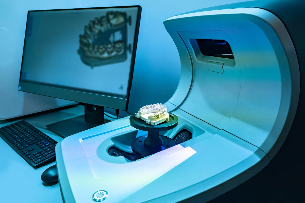
Indirect digitalisation involves scanning elastomeric impressions or plaster models with intraoral or extraoral (laboratory) scanners.
Another indirect digitalisation technique consists in scanning the impression material, if radiopaque, with Cone Beam Computed Tomography (CBCT), thus obtaining a digital model in DICOM format.
The literature on this topic includes several studies published in high-profile journals.
The study by Jelicich et al.
In a study conducted by Jelicich et al. (12), published in the Journal of Prosthetic Dentistry, scans obtained with laboratory scanners of polyvinylsiloxane (PVS) impressions showed greater accuracy in reproducing the spatial position of the abutments, compared to both direct intraoral scans and plaster models.
This evidence would indicate that the production of plaster models for full-arch impressions on natural teeth may not be necessary and that the conventional workflow could be simplified by digitalising the PVS impressions directly with a laboratory scanner.(12)
The study by Ender et al.
Results different from those obtained by Ender et al. (13) in which conventional vinyl polyether siloxane impressions showed significantly greater precision than intraoral scanners but equal accuracy.
This is probably due to the factors that influence intraoral scans which, in a large impression such as that of the full arch, can generate results that are not always repeatable.
However, no differences in precision or accuracy were found between scans of elastomer impressions and plaster models.
The study by Grande et al.
The study by Grande et al. (14), on a dental model, also highlighted better precision in the complete arch of scannable vinyl polysiloxane (Hydrorise Implant) compared to two intraoral scanners, with an accuracy that was also greater, though not significantly so.
However, these results lack comparison with scans of plaster models obtained from impressions.
Other contributions
Other authors have instead highlighted a minus in scanning impressions compared to scanning plaster models due to the reduced capacity of the scanner to capture deep hollow spaces in the contours and internal areas of the dental shape.(15)
It is true that scanning a negative with some undercut areas due to the dental shapes and slopes can be difficult and the resulting scan may not be perfect.
Therefore, in cases where the scanner is unable to correctly capture all the details in the dental shapes, it is definitely advisable to cast a plaster model and scan it to move to the digital workflow.(15)
Systems integration to improve clinical outcomes
Returning to the clinical aspect, however, it is true that creating a perfect impression with elastomeric materials in full-arch prosthetic rehabilitations is very challenging, even for the most expert professionals.
If the impression is not correct, it should be repeated until all the necessary details are captured along the entire arch.
The use of an intraoral scanner, allowing for easy partial scans to integrate any missing areas, could represent a very easy solution to obtain the requested information and avoid taking another impression.(12)
In this sense, integrating both systems could improve clinical outcomes. Furthermore, the ability of some radiopaque materials to be scanned with CBCT offers significant opportunities not only in the field of prosthetics, but also in guided surgery and integration between STL and DICOM files.
However, identifying the optimal radiographic settings for the CBCT scanner could reduce errors, thus representing a promising field of research for the future.
References
- Revilla-Leon M, Frazier K, da Costa JB, Kumar P, Duong ML, Khajotia S, et al. Intraoral scanners: An American Dental Association Clinical Evaluators Panel survey. The Journal of the American Dental Association. 2021 Aug 1;152(8):669-670.e2.
- Celeghin G, Franceschetti G, Mobilio N, Fasiol A, Catapano S, Corsalini M, et al. Complete-Arch Accuracy of Four Intraoral Scanners: An In Vitro Study. Healthcare. 2021 Mar 1;9(3):246.
- Revilla-León M, Jiang P, Sadeghpour M, Piedra-Cascón W, Zandinejad A, Özcan M, et al. Intraoral digital scans-Part 1: Influence of ambient scanning light conditions on the accuracy (trueness and precision) of different intraoral scanners. J Prosthet Dent. 2020 Sep;124(3):372–8.
- Syrek A, Reich G, Ranftl D, Klein C, Cerny B, Brodesser J. Clinical evaluation of all-ceramic crowns fabricated from intraoral digital impressions based on the principle of active wavefront sampling. Journal of Dentistry. 2010 Jul 1;38(7):553–9.
- Berrendero S, Salido MP, Valverde A, Ferreiroa A, Pradíes G. Influence of conventional and digital intraoral impressions on the fit of CAD/CAM-fabricated all-ceramic crowns. Clin Oral Invest. 2016 Dec 1;20(9):2403–10.
- Sailer I, Mühlemann S, Fehmer V, Hämmerle CHF, Benic GI. Randomized controlled clinical trial of digital and conventional workflows for the fabrication of zirconia-ceramic fixed partial dentures. Part I: Time efficiency of complete-arch digital scans versus conventional impressions. Journal of Prosthetic Dentistry. 2019 Jan 1;121(1):69–75.
- Revilla-León M, Subramanian SG, Att W, Krishnamurthy VR. Analysis of Different Illuminance of the Room Lighting Condition on the Accuracy (Trueness and Precision) of An Intraoral Scanner. Journal of Prosthodontics. 2021;30(2):157–62.
- Oh KC, Park J, Moon HS. Effects of Scanning Strategy and Scanner Type on the Accuracy of Intraoral Scans: A New Approach for Assessing the Accuracy of Scanned Data. Journal of Prosthodontics. 2020 Jul;29(6):518–23.
- Revilla-León M, Jiang P, Sadeghpour M, Piedra-Cascón W, Zandinejad A, Özcan M, et al. Intraoral digital scans: Part 2-influence of ambient scanning light conditions on the mesh quality of different intraoral scanners. J Prosthet Dent. 2020 Nov;124(5):575–80.
- Ferrari Cagidiaco E, Zarone F, Discepoli N, Joda T, Ferrari M. Analysis of The Reproducibility of Subgingival Vertical Margins Using Intraoral Optical Scanning (IOS): A Randomized Controlled Pilot Trial. J Clin Med. 2021 Mar 1;10(5):941.
- Bennani V, Aarts JM, Schumayer D. Correlation of pressure and displacement during gingival displacement: An in vitro study. J Prosthet Dent. 2016 Mar;115(3):296–300.
- Jelicich A, Scialabba R, Lee SJ. Positional trueness of abutments by using a digital die-merging protocol compared with complete arch direct digital scans and conventional dental impressions. J Prosthet Dent. 2024 Feb;131(2):293–300.
- Ender A, Mehl A. In-vitro evaluation of the accuracy of conventional and digital methods of obtaining full-arch dental impressions. Quintessence Int. 2015 Jan;46(1):9–17.
- Grande F, Celeghin G, Gallinaro F, Mobilio N, Catapano S. Comparison of the accuracy between full-arch digital scans and scannable impression materials: an in vitro study. Minerva Dent Oral Sc [Internet]. 2023 Apr [cited 2023 Jul 3]; Available from: https://www.minervamedica.it/index2.php?show=R18Y9999N00A23041706
- Bosniac P, Rehmann P, Wöstmann B. Comparison of an indirect impression scanning system and two direct intraoral scanning systems in vivo. Clin Oral Invest. 2019 May;23(5):2421–7.
Would you like more information about Zhermack Dental products and solutions?
Contact us


 Zhermack SpA has been one of the most important producers and international distributors of alginates, gypsums and silicone compounds for the dental sector for over 40 years. It has also developed solutions for the industrial and wellbeing sectors.
Zhermack SpA - Via Bovazecchino, 100 - 45021 Badia Polesine (RO), Italy.


