Full edentulism is a fairly common problem and is often treated will full removable dentures. Clinicians use different materials and techniques to take the final impression. Adopting the correct impression technique improves the quality of the restoration, and therefore improves the patient’s quality of life. (1)
Impression techniques for edentulous patients can be broken down into “one-stage” and “two-stage” techniques.
One-stage and two-stage techniques
One-stage techniques do not involve using a preliminary impression or a custom impression tray.
Two-stage techniques, on the other hand, involve taking a preliminary impression, which is used to make a gypsum model. This model is used to fabricate a custom impression tray, with which the final impression is taken. (2)
There is still a certain amount of confusion regarding the need to use a two-stage technique when taking impressions on edentulous patients, as, according to some studies, there are no patient-perceived differences following the fabrication of removable dentures. (3-5)
“Open-Mouth” and “Closed-mouth” techniques
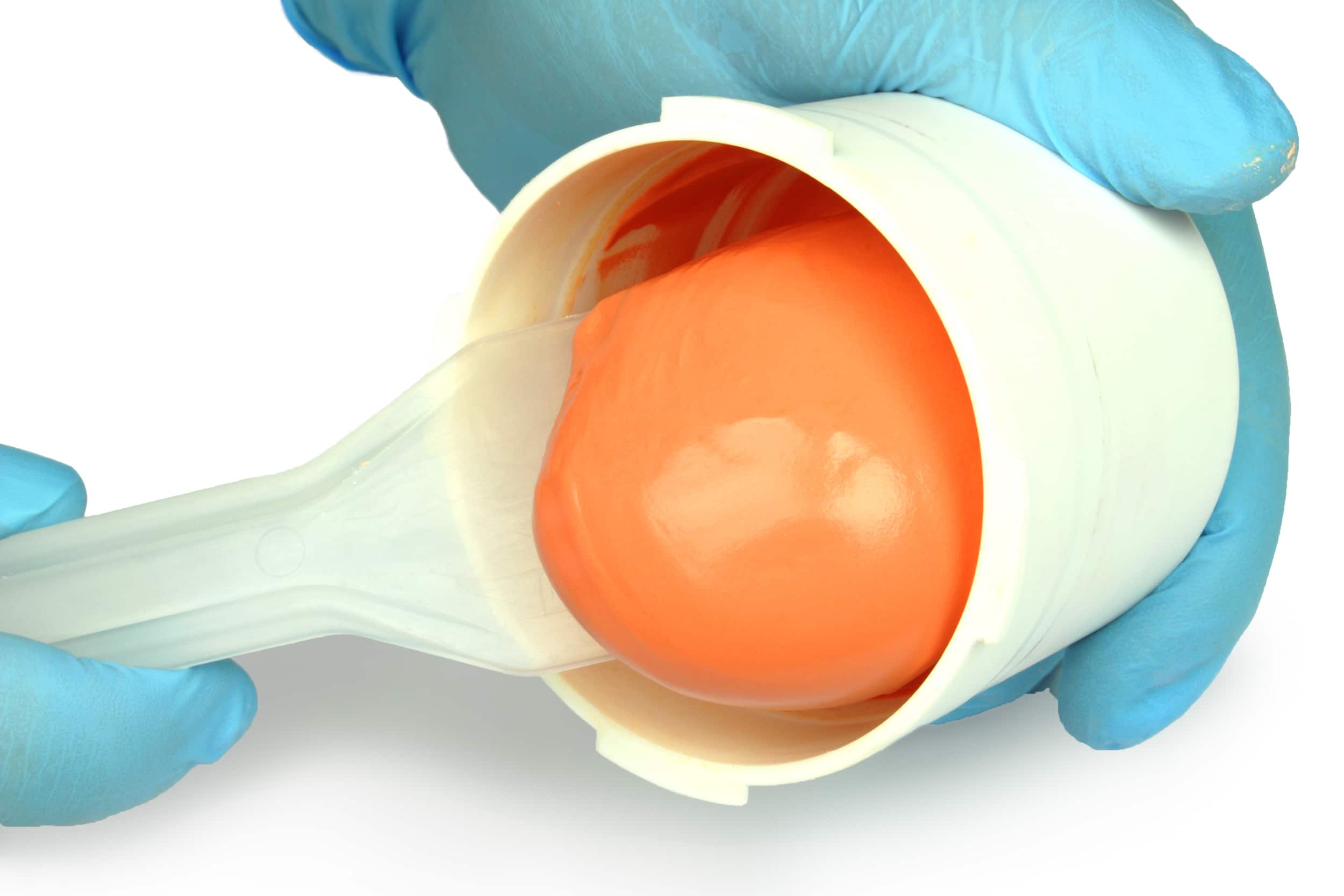
The secondary or final impression can be taken using “Open-mouth” or “closed-mouth” techniques (6), using one or more steps depending on whether the rim stage is included in the impression stage or is carried out previously. (7-10)
The rim stage consists in functionally or manually adapting certain materials on the edge of the impression tray, in order to create a peripheral seal between the impression tray and the tissues.
The rim stage is also defined as determining the extension of the prosthesis, by fitting the thermoplastic material around the marginal perimeter, by means of a functionalisation or manipulation of the tissues. (11)
In addition to this differentiation, there are several techniques for taking final impressions. (12)
Other techniques for taking final impressions
Over the years, the development of increasingly high-performance materials has resulted in the emergence and evolution of different techniques, which can be classified as: mucostatic, mucocompressive, selective pressure, functional and neutral zone techniques. (1)
The mucostatic technique
The mucostatic technique (13-15) aims to reproduce the soft tissues, while limiting the warping that can be caused by the material.
The anatomy of the patient is recorded statically, without in any way modifying the actual intraoral anatomical relationships. This technique is carried out using impression trays with special characteristics and low-density materials.
The mucocompressive technique
The mucocompressive technique (16,17) is based on the concept that, when chewing, the patient will apply a certain pressure to the tissues beneath the prosthesis and therefore the impression must be taken while applying a certain compression of the tissues.
However, restorations obtained with this type of impressions are imprecise and uncomfortable the rest of the time, as the tissues tend to exert a certain force on the prosthesis.
The selective pressure technique
The selective pressure technique (18,19) exploits the principle according to which certain areas of the edentulous crest are more likely to withstand masticatory loads without undergoing bone resorption processes, while others are at a greater risk.
According to this technique, the areas at a high risk of resorption are recorded in a completely static manner, whereas the pressure of the impression material is directed towards areas that can withstand the masticatory load.
Functional techniques
With functional techniques (14,20), while the impression is being taken, the patient is instructed to carry out a series of mimic or chewing movements or to pronounce certain letters, in order to properly functionalise the impression and adjust the extent of the future prosthesis also on the basis of functional movements.
The Neutral Zone technique
The neutral zone technique (21,22) aims to record the neutral zone, i.e. the space between the lips/cheeks and the tongue. The neutral zone is the space in which the patient’s prosthetic restorations will be located.
Want to find out more?
Discover the differences between preliminary and functional impressions for edentulous patients.
Establishing the technique and materials to be used
There is little literature evidence available regarding the technique of election when taking impressions for removable full dentures. (1)
Notwithstanding the results of certain studies, two-stage impressions are still the gold standard.
Establishing whether to use one or more materials and one or more techniques is therefore at the discretion of the clinician, who uses professional experience to choose the procedure that is most congenial to him/her, which is also often the most comfortable and affordable for the patient (23).
The ideal alginate for impressions on edentulous patients
Zhermack offers a vast range of alginates with characteristics that are suitedto use in partly or fully edentulous patients for the fabrication of partial and full removable dentures.
More specifically, Neocolloid is an alginate with a long setting time, designed for obtaining adequate impressions of the mucous membranes of edentulous patients and that, thanks to its physical and chemical characteristics, allows an excellent reproduction of the maxillary mucosae.
References
- Jayaraman, S., Singh, B. P., Ramanathan, B., Pillai, M. P., MacDonald, L., & Kirubakaran, R. (2018). Final‐impression techniques and materials for making complete and removable partial dentures. Cochrane Database of Systematic Reviews, (4).
- Trapozzano VR. Securing edentulous impressions with zinc oxide-eugenol impression paste. Journal of the American Dental Association 1939;26(9):1527-31.
- Komagamine Y, Kanazawa M, Sato Y, Iwaki M, Jo A, Minakuchi S. Masticatory performance of different impression methods for complete denture fabrication: A randomized controlled trial. J Dent 2019;83:7–11. https://doi.org/10.1016/j.jdent.2019.01.009.
- Albuquerque IS, Regis RR, de Souza RF, Gurgel KF, Silva PG, Pinto-Fiamengui LMS, et al. Is a two-step impression mandatory for complete denture fabrication on the severely resorbed mandible? A randomized trial on patient perception and denture quality. Journal of Dentistry 2020;98:103356. https://doi.org/10.1016/j.jdent.2020.103356.
- Hyde TP, McCord JF. Survey of prosthodontic impression procedures for complete dentures in general dental practice in the United Kingdom. The Journal of Prosthetic Dentistry 1999;81:295–9. https://doi.org/10.1016/S0022-3913(99)70272-6.
- Boucher CO. A critical analysis of mid-century impression techniques for full dentures. Journal of Prosthetic Dentistry 1951;1(4):472-91.
- Loh PL. An alternative for making master impressions for complete dentures. Journal of the American Dental Association 1997;128(10):1436-7.
- Minagi S, Sato Y, Akagawa Y, Tsuru H. Concept and technique for making an accurate final impression for complete dentures using a thixotropic impression material. International Journal of Prosthodontics 1987;1(2):149-52.
- ChaBee NR, Cooper LF, Felton DA. A technique for border molding edentulous impressions using vinyl polysiloxane material. Journal of Prosthodontics 1999;8(2):129-34.
- Smith DE, Toolson LB, Bolender CL, Lord JL. One-step border molding of complete denture impressions using a polyether impression material. Journal of Prosthetic Dentistry 1979;41(3):347 51.
- Ferro KJ, Morgano SM, Driscoll CF, Freilich MA, Guckes AD, Knoernschild KL, et al. The glossary of prosthodontic terms – ninth edition. Journal of Prosthetic Dentistry May 2017;117(5S):e1-105.
- StarckeENJr. A historical review of complete denture impression materials. Journal of the American Dental Association 1975;91(5):1037-41.
- AddisonPI. Mucostatic impressions. Journal of the American Dental Association 1944;31(13):941-6.
- Petropoulos, V. C., & Rashedi, B. (2003). Current concepts and techniques in complete denture final impression procedures. Journal of Prosthodontics, 12(4), 280-287.
- Bindhoo, Y. A., Thirumurthy, V. R., & Kurien, A. (2012). Complete mucostatic impression: a new attempt. Journal of Prosthodontics: Implant, Esthetic and Reconstructive Dentistry, 21(3), 209-214.
- Antonelli, J., Guerrero, M., Georgescu, M., & Ortiz, J. (2019). Quantifying flabby ridge tissue displacement during impression-making for patients with combination syndrome. Compend Contin Educ Dent, 40(8).
- Alqattan, W. A., Alalawi, H. A., & Khan, Z. A. (2016). Impression techniques and materials for complete denture construction. Dent Health Curr Res 2, 1, 13-17.
- Gupta, A., Singhal, P., & Negi, P. (2014). Selective pressure impression technique: an overview. Journal of Evolution of Medical and Dental Sciences, 3(29), 8110-8115.
- Duncan, J. P., Raghavendra, S., & Taylor, T. D. (2004). A selective-pressure impression technique for the edentulous maxilla. The Journal of prosthetic dentistry, 92(3), 299-301.
- Kršek, H., & Dulčić, N. (2015). Functional impressions in complete denture and overdenture treatment. Acta stomatologica Croatica: International journal of oral sciences and dental medicine, 49(1), 45-53.
- Makzoumé, J. E. (2004). Morphologic comparison of two neutral zone impression techniques: A pilot study. The Journal of prosthetic dentistry, 92(6), 563-568.
- Gahan, M. J., & Walmsley, A. D. (2005). The neutral zone impression revisited. British dental journal, 198(5), 269-272.
- Tsirogiannis P, Neophytou S, Reul A, Heydecke G, Reissmann DR. Can we measure patients’ perception during dental impressions? The Burdens in Dental Impression-Making Questionnaire – BiDIM-Q. Journal of Prosthodontic Research 2017;61:34–42. https://doi.org/10.1016/j.jpor.2016.03.003.
Do you want more information on Zhermack Dental products and solutions?
Contact us


