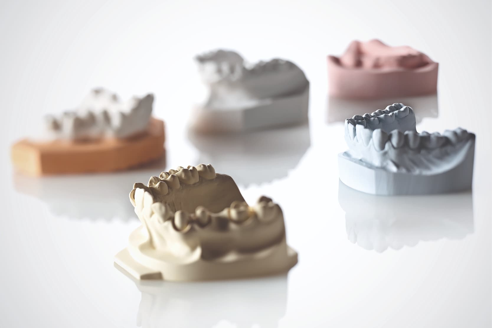
A model is a positive replica of the dentition and surrounding structures that is used for the diagnostic study of the situation and as a basis for preparing prosthetic and orthodontic appliances.[1]
There are study models, also known as diagnostic or preliminary models, working models and master models.[2]
Models intended for various uses must offer different physical and mechanical properties.
Characteristics and purposes of orthodontic models
Study/preliminary/diagnostic models are used for studying the case or preparing a custom impression tray with which to make a master model, which is the model on which the prosthetic product will actually be fabricated. Working models, on the other hand, are used to carry out certain technical steps.
These differences imply different needs.
When analysing a clinical case with a study/diagnostic/preliminary model, absolute accuracy is not required, because the model is merely used for preliminary diagnostic purposes. This means that materials that are simpler and faster to use and offer a lower level of accuracy can be preferred.
If the model is to be used to prepare a permanent crown, on the other hand, utmost accuracy is required, and we will use a master model with high surface detail reproduction and dimensional stability characteristics.
Another characteristic a model must possess, depending on the processing steps it will undergo, is refractoriness, i.e. the ability to withstand high temperatures. It should be pointed out that in all traditional “lost wax” techniques, models are subjected to very high temperatures.
Then when talking about models, an important distinction must be made depending on the preparation method used.
Model preparation methods
Models can be prepared using classic/analogue methods (in which the model is cast) or with CAD-CAM/digital methods (3D printing or milling). [3.4]
Classic/analogue method
This preparation method is that most commonly used and the longest-standing in dental practice. It always requires a physical negative mould of the intraoral structures of interest, i.e. an impression, which must possess physical and mechanical characteristics such as to allow the casting inside it of a fluid material, which, as it sets, will form the positive, i.e. the exact replica of the intraoral structures, in other words, the model.
The impression material must therefore be compatible with the material to be cast inside it, offer good wettability and reduce the expansion or contraction of the model material during setting.[5]
Using dental gypsums
The model materials that can be used with a traditional technique (casting) consist of dental gypsums and model resins.[6]
Dental gypsums are obtained by calcination and there are β (beta) gypsums, from which type I gypsums and type II gypsums are obtained, and α (alpha) gypsums from which type III gypsum is obtained; if additives are added to α (alpha) gypsums, type IV, improved type IV and type V gypsums can also be obtained.
The various types have different characteristics and indications.[7]
- Type I: soft impression gypsum, also known as plaster of Paris.
- Type II: soft model gypsum, suitable for study models or for model assembly on an articulator
- Type III: hard gypsum, for making highly-resistant models
- Type IV: extra-hard gypsum, suitable for making fixed prostheses, offering high resistance and low expansion
- Type V: extra-hard gypsum, suitable for making fixed prostheses, offering high resistance and low expansion
The expansion of the gypsum is a fundamental characteristic, since it will compensate any contractions of the material to be made on the model.
Want to find out more? Discover the comparison between gypsum models and 3D models that can be printed
If, for example, we want to make the metal substructure of a removable prosthesis with a non-noble metal alloy characterised by significant shrinkage, we should choose a type V gypsum with high expansion in order to compensate the contraction of the metal.
Choosing resin materials
Besides gypsum, the most commonly-used material, we can also use resin materials to cast a model, the most common types being[8.10]:
- epoxy resin;
- polyurethane resin.
Other materials have been proposed with the aim of obtaining more accurate and resistant models, such as resin-metal composites[11], resin-modified gypsums[12], reinforced polyurethane resins[13] and electrodeposition models[14].
CAD-CAM/digital method
The methods for fabricating a model using CAM (Computer Aided Manufacturing) technology can be broken down into two main families: 3D printing and milling.[4]
Each of these two methods has advantages and disadvantages in terms of management costs, material wastage and final accuracy[15-17].
In both cases the most commonly-used material is polymethyl methacrylate or PMMA; in the milling method discs of PMMA are used, whereas in 3D printing it is cured layer after layer until the final model is formed.[4]
In 3D printing, polypropylene, polycarbonate, polystyrene, acrylonitrile butadiene styrene, etc… can also be used[18].
Whereas gypsum materials tend to express a certain degree of expansion during the setting phase, resin materials, on the other hand, tend to contract. This behaviour should be taken into account during 3D printing and measures must be taken to limit this phenomenon and avoid introducing imprecision.
Different types of resin are used and there is, potentially, a broad spectrum of materials that can be employed to make models using CAD-CAM/digital technologies, depending on the accuracy and resistance desired.
References
[1] https://www.dentisti-italia.it/glossario/638_modello-dentale.html
[2] https://online.scuola.zanichelli.it/labodonto/files/2018/04/Mappa_Ud9.pdf
[3] Cicciù, M., Fiorillo, L., D’Amico, C., Gambino, D., Amantia, E. M., Laino, L., … & Cervino, G. (2020). 3D digital impression systems compared with traditional techniques in dentistry: A recent data systematic review. Materials, 13(8), 1982.
[4] Jeong, Y. G., Lee, W. S., & Lee, K. B. (2018). Accuracy evaluation of dental models manufactured by CAD/CAM milling method and 3D printing method. The journal of advanced prosthodontics, 10(3), 245-251.
[5] Ragain, J. C., Grosko, M. L., Raj, M., Ryan, T. N., & Johnston, W. M. (2000). Detail reproduction, contact angles, and die hardness of elastomeric impression and gypsum die material combinations. International Journal of Prosthodontics, 13(3).
[6] https://online.scuola.zanichelli.it/labodonto/files/2018/04/Mappa_Ud6_1.pdf
[7] No, A. A. S. (2000). Dental gypsum products. Chicago: American dental association council on dental materials and equipment.
[8] Chaffee, N. R., Bailey, J. H., & Sherrard, D. J. (1997). Dimensional accuracy of improved dental stone and epoxy resin die materials. Part II: Complete arch form. The Journal of prosthetic dentistry, 77(3), 235-238.
[9] Niekawa, C. T., Kreve, S., A’vila, G. B., Godoy, G. G., da Silva, J. E. V., & Dias, S. C. (2017). Analysis of the mechanical behavior and surface rugosity of different dental die materials. Journal of International Society of Preventive & Community Dentistry, 7(1), 34
[10] Derrien, G., & Sturtz, G. (1995). Comparison of transverse strength and dimensional variations between die stone, die epoxy resin, and die polyurethane resin. The Journal of prosthetic dentistry, 74(6), 569-574.
[11] Fan, P. L., Powers, J. M., & Reid, B. C. (1981). Surface mechanical properties of stone, resin, and metal dies. Journal of the American Dental Association (1939), 103(3), 408-411.
[12] Duke, P., Moore, B. K., Haug, S. P., & Andres, C. J. (2000). Study of the physical properties of type IV gypsum, resin-containing, and epoxy die materials. The Journal of prosthetic dentistry, 83(4), 466-473.
[13] Black, E. M. E. (2015). Polyurethane research for applications in the field of dentistry: Limiting side reactions in monomer development and synthesizing N-capped polymenthide.
[14] Stackhouse Jr, J. A. (1980). Electrodeposition in dentistry. A review of the literature. The Journal of Prosthetic Dentistry, 44(3), 259-263.
[15] Van Noort, R. (2012). The future of dental devices is digital. Dental materials, 28(1), 3-12.
[16] Yau, H. T., Yang, T. J., & Lin, Y. K. (2016). Comparison of 3-D Printing and 5-axis Milling for the Production of Dental e-models from Intra-oral Scanning. Computer-aided design and applications, 13(1), 32-38.
[17] Kasparova, M., Grafova, L., Dvorak, P., Dostalova, T., Prochazka, A., Eliasova, H., … & Kakawand, S. (2013). Possibility of reconstruction of dental plaster cast from 3D digital study models. Biomedical engineering online, 12(1), 1-11.
[18] Jockusch, J., & Özcan, M. (2020). Additive manufacturing of dental polymers: Dental Materials Journal, 2019-123.
Do you want more information on Zhermack Dental products and solutions?
Contact us
: the principles of dental preparation")


