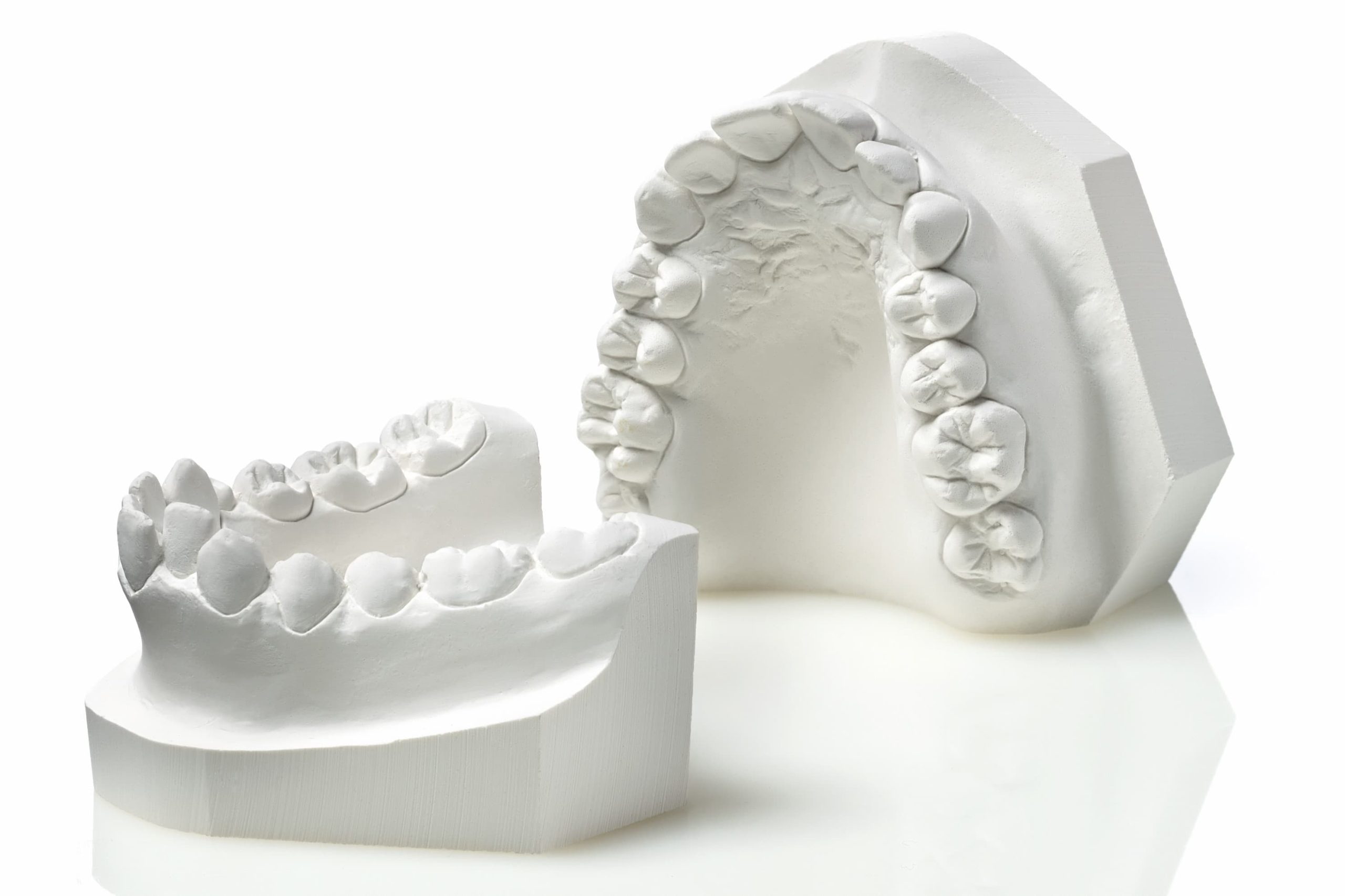
Dental models are positive reproductions of a patient’s intraoral situation. (1)
They can be fabricated using either the conventional technique, by casting gypsum inside an impression, or a CAD/CAM digital technique, by milling or 3D printing. (2)
Depending on how they are used, Models can be divided into two families (1, 3, 4):
- Study or Diagnostic Models
- Working or Master Models
Let’s focus on Study Models.
Study or Diagnostic Models
Unlike Working Models (which have a purely practical value and form the base on which the dental technician can fabricate the device, be it a crown, a bridge, a removable denture, or an orthodontic appliance, etc.), Study Models are not used to produce any prosthesis or appliance.
When used at the diagnostic stage or to choose the most suitable treatment plan, they are valuable aids for complex prosthetic cases. (5, 6)
However, Study Models play a fundamental role in a specific branch of dentistry: orthodontics.
Study Models in Orthodontics
In orthodontics, Study Models constitute an essential part of the diagnostic records (7). In order to carry out a proper “case study”, orthodontists rely primarily on information from three sources: photographs, X-rays and Study Models. (8, 9)
In certain special circumstances, the practitioner might also ask for second-level imaging procedures, like CBCT, anteroposterior teleradiographs, MRIs, etc. (10)
Why are gypsum Models so useful for diagnosis in orthodontics?
First and foremost, even the most attentive observer would be unable to capture all the information that a thorough analysis of stone Models can provide.
Being able examine the arches and the relationships between them from a convenient extraoral perspective helps the dentist to better understand the patient’s malocclusion type and to choose the most suitable treatment plan.
Being able to directly observe the interocclusal situation in the three planes of space provides the orthodontist with important information on any discrepancies on the transverse, vertical and sagittal planes.
Not only does it make it possible to identify certain space problems, it also enables their objective measurement.
Observation of the interocclusal situation in the three planes of space
On the transverse plane, Study Models can be used to measure the width of the palate and any discrepancy in relation to the width of the mandibular arch, and consequently establish the correct magnitude of its expansion. (11)
Knowing this relationship is of paramount importance when using a rapid palatal expander to plan the number of activations required to normalise the palatal contraction.
On the vertical plane, it is possible to measure the entity of the overbite, i.e. the vertical discrepancy in the anterior segment. (12)
Sagittally, it is possible to establish the severity of a class II or III malocclusion, by physically measuring the discrepancy. This finding can guide the dentist towards the right type of treatment.
In addition to the measurements resulting from the relationship between the arches, there is another fundamental type of assessment: space analysis.
Space analysis
Space analysis (13) is the relationship between the space required and the space available in the arch for the alignment of the teeth.
This analysis is carried out by measuring the mesiodistal size of each tooth using a caliper and comparing the sum of all of these measurements with the length of the arch perimeter on the bone crest.
Space analysis provides an objective quantification of crowding, information that is essential for diagnosis and consequently for the treatment plan.
There must be a certain ratio between the sum of the mesiodistal sizes of the upper arch and the sum of the mesiodistal sizes of the lower arch.
This ratio is known as the “Bolton Index“. (14) If the Bolton index is abnormal, occlusion will always have a certain degree of imperfection.
In addition to measuring mesiodistal tooth size, stone models can also be used to measure the depth of the curve of Spee and therefore obtain further information on the extent of crowding. (15)
The Zhermack solution for fabricating stone models
Having examined the entire series of diagnostic data recorded by analysing the Study Models, it goes without saying that without this kind of record it is difficult, if not impossible, to obtain a correct and precise orthodontic diagnosis and prepare a treatment plan suited to the patient.
Zhermack offers several high-performance products for the fabrication of stone Models. Elite Dental Stones is the wide range of Zhermack stones, with low expansion even after 48 hours, able to satisfy the diverse requirements of dental technicians.
With its type 3 and type 4 gypsums, Elite Dental Stones it provides specific and separate solutions, ranging from the fabrication of antagonist or diagnostic Models to the development of master models.
Elite Ortho is the Zhermack solution designed for the fabrication of orthodontic study models.
Bibliography
- The Glossary of Prosthodontic Terms: Ninth Edition. J Prosthet Dent 2017;117: e1-e105 n.d.
- Jeong, Y. G., Lee, W. S., & Lee, K. B. (2018). Accuracy evaluation of dental models manufactured by CAD/CAM milling method and 3D printing method. The journal of advanced prosthodontics, 10(3), 245-251.
- Rudd, K. D. (1968). Making diagnostic casts is not a waste of time. The Journal of Prosthetic Dentistry, 20(2), 98-100.
- Solow, R. A. (2016). Quantified diagnostic work-up casts: applications for interdisciplinary treatment planning. General Dentistry, 64(3), 37-46.
- Amin, K., Vere, J., Thanabalan, N., & Elmougy, A. (2019). Occlusal concepts and considerations in fixed prosthodontics. Primary Dental Journal, 8(3), 20-27.
- Abduo, J. (2012). Virtual prosthodontic planning for oral rehabilitation: a pilot study. In Proceedings of the Workshop on New Trends of Computational Intellegence in Health Applications (pp. 34-42).
- Rischen, R. J., Breuning, K. H., Bronkhorst, E. M., & Kuijpers-Jagtman, A. M. (2013). Records needed for orthodontic diagnosis and treatment planning: a systematic review. PLoS one, 8(11), e74186.
- Jackson, T. H., Kirk, C. J., Phillips, C., & Koroluk, L. D. (2019). Diagnostic accuracy of intraoral photographic orthodontic records. Journal of Esthetic and Restorative Dentistry, 31(1), 64-71.
- Paredes, V., Gandia, J. L., & Cibrián, R. (2006). Digital diagnosis records in orthodontics. An overview. Med Oral Patol Oral Cir Bucal, 11(1), E88-93.
- Hechler, S. L. (2008). Cone-beam CT: applications in orthodontics. Dental Clinics of North America, 52(4), 809-823.
- Eslami Amirabadi, G., Golshah, A., Derakhshan, S., Khandan, S., Saeidipour, M., & Nikkerdar, N. (2018). Palatal dimensions at different stages of dentition in 5 to 18-year-old Iranian children and adolescent with normal occlusion. BMC oral health, 18(1), 1-6.
- Agrawal, G. (2016). Deep bite its etiology, diagnosis and management: a review. Journal of Orthodontics, 2(4), 12.
- Fleming, P. S., Marinho, V., & Johal, A. (2011). Orthodontic measurements on digital study models compared with plaster models: a systematic review. Orthodontics & craniofacial research, 14(1), 1-16.
- Bailey, L. A. (1998). The Bolton analysis revisited.
- Marshall, S. D., Caspersen, M., Hardinger, R. R., Franciscus, R. G., Aquilino, S. A., & Southard, T. E. (2008). Development of the curve of Spee. American Journal of Orthodontics and Dentofacial Orthopedics, 134(3), 344-352.
Do you want more information on Zhermack Dental products and solutions?
Contact us


