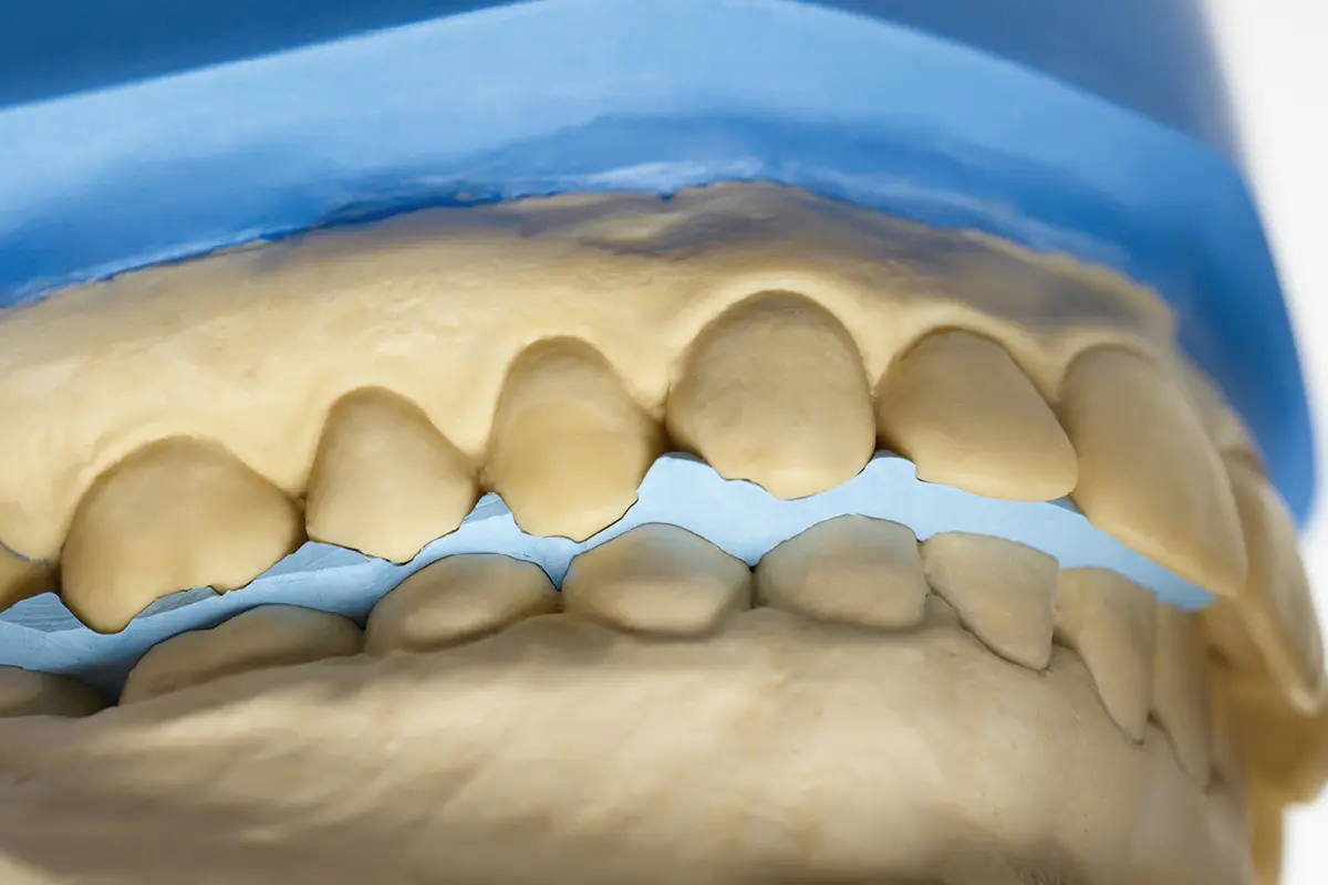
Scannable bite registration materials are extremely useful in clinical dental practice, allowing the accurate transfer of the maxillomandibular relationship and offering stability to the dental models mounted in the articulator.
Advantages of occlusion materials
The scientific literature highlights several important roles played by occlusal registration materials, in particular in terms of possible advantages that they can provide the clinician and the dental technician.
Let’s take a closer look.
Transfer of the maxillomandibular relationshipfrom the intraoral environment to the mechanical articulator
The first advantage consists in the transfer of the maxillomandibular relationshipfrom the patient’s intraoral environment to the mechanical articulator in the absence of a stable relationship in maximum intercuspation (1,2).
This allows dental technicians to work on articulator-mounted dental models, which attempt to replicate the mutual position of the patient’s upper and lower teeth.
Horizontal and vertical stability
The second advantage is represented by the horizontal and vertical stabilityprovided to the models during assembly in the articulator (3).
This is a key objective of bite registration, and is guaranteed by only the registration material.
It prevents horizontal rotation, translation, or roto-translation of the dental models during mounting, while providing the basis for planning and manufacturing restorations.
Positioning of the models according to defined occlusal criteria
Another advantage is that it makes it possible to model the teeth according to well-defined occlusal criteria.
The precise positioning of the models in the articulator implies a certain dental shape to be given to the restorations in relation to the antagonist.
Furthermore, the technician can effectively evaluate the prosthetic space available and define the correct thicknesses of the materials to be used (4).
Reduction of intraoral occlusal adaptations
Accurate bite registration can reduce the need to make significant intraoral modifications to prosthetic restorations.
The result is a reduced need for occlusal adjustments during the clinical phase, consequently also reducing the time necessary for adjustments, and a reduced risk of structural and aesthetic weakening of denture materials (particularly ceramics) (5).
Analysis of post-repositioning occlusal relationships
Yet another advantage is given by the possibility of carrying out a detailed analysis of the occlusal relationships following repositioning in the articulator.
In cases where, for example, it is necessary to carry out multiple and extensive occlusal adjustments (for example full-arch rehabilitations on implants), the clinician can decide to detect the occlusion with the appropriate registration materials and have the models repositioned in the articulator for the technician in the most appropriate way in order to carry out the necessary adjustments and polish the prosthesis before returning it.
Useful information on occlusal thicknesses
The use of occlusal registration materials allows us to understand the thicknesses of the prosthetic materials at the occlusal level.
When trying the prosthetic frameworks in the mouth, before applying the coating materials, the clinician can decide to place some occlusal registration material on the restoration in order to provide the dental technician with useful information regarding both the articulation and the residual occlusal thickness (6,7).
Subsequent control in case of injections
The professional has the possibility of checking the occlusion a posteriori in cases of intramuscular or intra-articular injections (8).
The uses of scannable material for bite registration
As anticipated in the introduction, these just listed above are just some of the advantages that occlusion materials can provide the clinician and dental technician.
Today, thanks in particular to the advent of digital techniques, if they are scannable using intraoral or extraoral scanners, these materials can be useful for transferring all this information in the digital environment as well (9,10).
A very quick use of scannable material for bite registration is to transfer the mutual position of the jaws once they have also been scanned.
This procedure can be particularly useful in partially edentulous patients, since in the absence of dental elements fully digital detection of the occlusion can be inaccurate (11).
And by broadening the concept to patients who are entirely edentulous or waiting to be rehabilitated with implant prostheses, scannable materials can also be useful to indicate what will be the vertical dimension of the patient (12,13).
Another very interesting use involves the use of scannable materials in association with the facial bow in order to transfer the correct position of the upper jaw onto the digital articular (9).
Want to know more?
Find out how to choose the material for bite registration
In all these cases, however, it is necessary to pay particular attention to how materials are used and also how all components are integrated into the digital workflow in order to obtain predictable results in clinical practice.
In fact, in many of these applications, occlusal registration materials are used in unusual clinical conditions that do not strictly reflect their specific indications for use.
When they are used to articulate the occlusion starting from toothed arches instead, their accuracy and precision in determining the position of the contacts is well demonstrated by the literature, even going so far as to locate occlusal contact areas spaced up to 50 μm apart (14).
Conclusions
As mentioned earlier, scannable bite registration materials are extremely useful materials in clinical dental practice, allowing the accurate transfer of the maxillomandibular relationship and offering stability to the dental models mounted in the articulator.
Furthermore, thanks to their scannability, they can be integrated into the digital workflow to facilitate procedures such as the repositioning of the jaws and the determination of the vertical dimension in partially and fully edentulous patients.
However, it is essential to carefully consider the specific applications and couplings in the digital workflow in order to ensure predictive results in clinical practice.
References
- The Glossary of Prosthodontic Terms. The Journal of Prosthetic Dentistry. 2017 May;117(5):C1-e105.
- Freilich MA, Altieri JV, Wahle JJ. Principles for selecting interocclusal records for articulation of dentate and partially dentate casts. J Prosthet Dent. 1992 Aug;68(2):361–7.
- D KP, Prasad BR, D AP, Mehra D. INTEROCCLUSAL RECORDS IN PROSTHODONTIC REHABILITATIONS – MATERIALS AND TECHNIQUES – A LITERATURE REVIEW. Journal of Health and Allied Sciences NU. 2012 Sep;02(3):54–60.
- Darroudi M, Ariens ZPA, Zinsmeister VZ, Breuning KH. [To bite or to scan? Dental impressions with alginate, PVS or -intra-oral scanning; processing time and patient comfort. A pilotstudy]. Ned Tijdschr Tandheelkd. 2017 Feb;124(2):91–5.
- Herpel C, Rammelsberg P, Rues S, Zenthöfer A, Seceleanu I, Corcodel N. Color stability of individually stained monolithic zirconia following occlusal adjustment. Journal of Esthetic and Restorative Dentistry. 2021;33(2):387–93.
- Vidotti HA, Pereira JR, Insaurralde E, de Almeida ALPF, do Valle AL. Thermo and mechanical cycling and veneering method do not influence Y-TZP core/veneer interface bond strength. J Dent. 2013 Apr;41(4):307–12.
- Lin WS, Ercoli C, Feng C, Morton D. The Effect of Core Material, Veneering Porcelain, and Fabrication Technique on the Biaxial Flexural Strength and Weibull Analysis of Selected Dental Ceramics. Journal of Prosthodontics. 2012;21(5):353–62.
- Mobilio N, Catapano S. Effect of experimental jaw muscle pain on occlusal contacts. J Oral Rehabil. 2011 Jun;38(6):404–9.
- Petre A, Drafta S, Oancea L. A new technique to transfer the upper maxillary arch position using a facebow, a transfer table, and a reference block with a CAD application. J Prosthodont. 2024 Feb;33(2):195–200.
- Li J, Sommer C, Wang HL, Lepidi L, Joda T, Mendonca G. Creating a virtual patient for completely edentulous computer-aided implant surgery: A dental technique. J Prosthet Dent. 2021 Apr;125(4):564–8.
- Ren S, Morton D, Lin WS. Accuracy of virtual interocclusal records for partially edentulous patients. J Prosthet Dent. 2020 Jun;123(6):860–5.
- Srinivasan M, Kalberer N, Naharro M, Marchand L, Lee H, Müller F. CAD-CAM milled dentures: The Geneva protocols for digital dentures. The Journal of Prosthetic Dentistry. 2020 Jan 1;123(1):27–37.
- Patil PG, Nimbalkar-Patil S. Maxillomandibular relationship record for complete arch/mouth implant restorations using putty-elastomeric occlusion rim at healing abutment level. Contemp Clin Dent. 2015;6(3):318–20.
- Rovira-Lastra B, Khoury-Ribas L, Flores-Orozco EI, Ayuso-Montero R, Chaurasia A, Martinez-Gomis J. Accuracy of digital and conventional systems in locating occlusal contacts: A clinical study. J Prosthet Dent. 2023 Aug 21;S0022-3913(23)00481-X.
Would you like more information about Zhermack Dental products and solutions?
Contact us


