A full removable denture is one of the most complex and diverse treatments that can be encountered in clinical and technical settings (1–3).
The differences between completely edentulous patients, the different types of osteo-mucosal resorption that can occur and the incongruence between the maxillary bones and neuromuscular control make treatment with full removable dentures extremely individual and complex, for both clinicians and dental technicians (4,5).
Several analogue workflows can be used to fabricate a full removable denture (4,6). In order to simplify the context, however, these procedures can be considered as more or less extreme variants of the conventional linear technique (7).
The 9 steps of the conventional linear technique
This technique, as has already been extensively described (7), consists of a total of 9 steps (5 clinical and 4 technical steps):
- the edentulous patient’s first appointment and taking of preliminary alginate impressions of the arches;
- development of the preliminary gypsum models and fabrication of the custom impression trays;
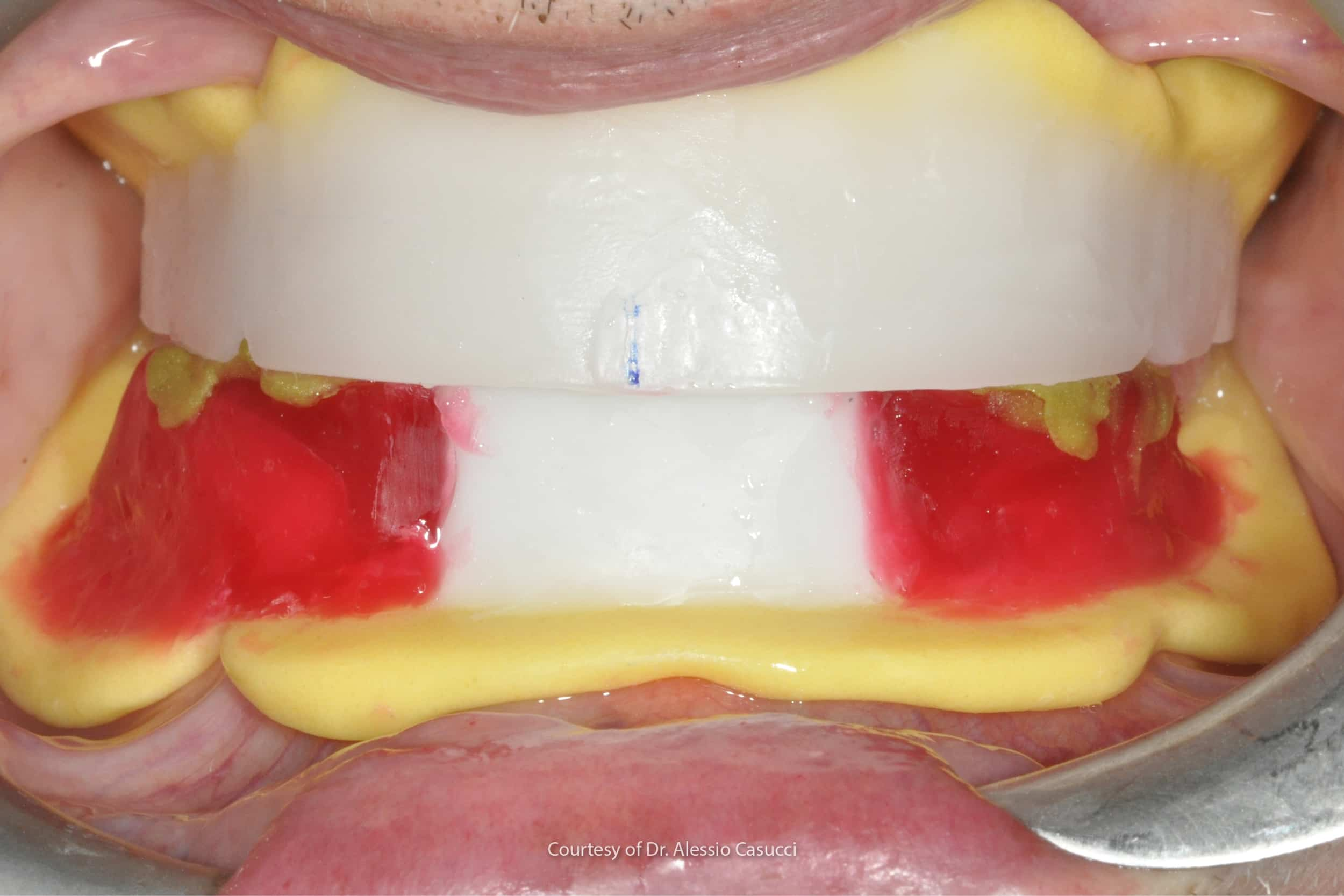
- functional secondary impressions;
- development of the master models and fabrication of the occlusion rims;
- recording of the centric relation;
- mounting of the models with the occlusion rims on an articulator, choice and assembly of the frontal elements and diatorics;
- aesthetic, phonetic and functional testing and patient consent;
- finishing of the dentures in the laboratory;
- delivery of the denture to the patient following a fit check.
In the first clinical step an in-depth history, both medical and dental, must be collected in order to better characterise the clinical case. It is, however, also necessary to understand the patient’s psychological status and expectations.
The practitioner-edentulous patient relationship
As we know from the scientific literature, edentulous patients can be classified into different types, depending on the psychological impact of the situation of edentulism and rehabilitation with a removable denture (2).
In this sense, the practitioner-patient relationship is fundamental, because the patient’s ability to adjust to the new situation with the removable denture is not necessarily simple (8). Patients are therefore likely to benefit from a good trusting relationship with their dentist (2).
Extra/intraoral examinations and x-rays
After this aspect, which is perhaps the most important with removable dentures, the dentist proceeds first with an extraoral physical exam, and then with an intraoral exam, in order to continue monitoring the health of the soft tissues and bone crests and take appropriate x-rays (OPT, intraoral x-rays), photographs and preliminary impressions (7).
Preliminary impressions with Schreinemaker impression trays
Preliminary impressions must be panoramic and cover the whole arch.
They must reproduce the whole of the basal area of the edentulous maxillae, the extent of the vestibular fornices and the anatomical elements that will support the future denture (canine protrusion, maxillary tuberosities, alveolar ridges, etc.).
These impressions are generally taken using alginate and with an impression tray that is either commercially available or custom made for the edentulous patient (Schreinemaker) (7).
Schreinemaker impression trays come in an assortment of 10 shape variants (5 upper + 5 lower) and with a measuring compass. They are always perforated, have a riveted edge and reflect the anatomy of edentulous patients, making them particularly advantageous for reducing alginate thicknesses.
It is the clinician’s duty to decide which type of impression tray to use, based on the degree of bone resorption and the residual anatomy.
Secondary or functional impressions
Once the preliminary models have been created and the custom impression trays fabricated, the clinician proceeds to the second clinical step, i.e. that of the secondary or functional impressions, the purpose of which is to record the patient’s muscle dynamics (6,7).
The muscles must be recorded in the impression so that the edge of the denture is in perfect harmony with the muscle function.
The success of the secondary impression in any case depends greatly on the custom impression tray and the clinical rim stage.
The clinical phase, therefore proceeds with a check on the adjustment of the custom impression tray, peripheral rimming with thermoplastic or elastomeric materials by functionalising the muscles and frenula and, lastly, the taking of an impression of the tissues (6,7).
Materials for secondary impressions for full dentures
The materials used to take secondary impressions for full dentures varies greatly.
In the United States and Saudi Arabia, polyvinylsiloxanes appear to be the materials most commonly used to take secondary impressions of edentulous maxillae (9–11), whereas in other countries, such as India and Nepal, zinc oxide-eugenol remains the material of choice (12,13).
It should be pointed out that many clinicians also use other materials for secondary impressions, such as polyethers and polysulphides (9–13). This depends on the technique used and the school of thought followed, which also dictates the type of custom impression tray to be made, not merely the technique to be carried out.
Recording the patient’s vertical dimension and the centric relationship
The third clinical step, on the other hand, consists in recording the patient’s vertical dimension (14) and the centric relationship, using resin plates and wax occlusion rims.
This is another very important step, since the patient’s vertical dimension is crucial for proper rehabilitation.
Furthermore, in this clinical step, the prosthetic space of the upper and lower arches and the spatial orientation of the occlusal plane will be defined and the neutral area in which the technician will position the teeth will be established (7).
Once again clinicians can potentially use different techniques, which vary depending on personal preferences and schools of thought,by carrying out, for example, aesthetic and phonetic tests, etc.
Aesthetic, phonetic and functional tests
Once the intermaxillary relations have been defined, the aesthetic, phonetic and rim function tests are carried out, with the teeth positioned by the dental technician.
During this step, the clinician assesses the actual performance of the occlusion rims with the teeth.It is therefore possible to make changes to the position of the teeth depending on phonetics, masticatory function, functional stability, but also in relation to the patient’s aesthetic expectations (7).
In some cases the clinician may decide to carry out a first test with the front teeth (canine to canine) alone, and then carry out a second test including the premolars and molars, in accordance with the school of Pound (15).
Fabricating full dentures
Once the clinical assessment of the placement of the teeth has been completed and the patient’s consent has been obtained, the actual fabrication of the full denture can take place.
Depending on the technique chosen by the dentist, the denture can be fabricated analogically using conventional flasking techniques. These techniques traditionally involve the use of addition silicones such as Elite Double and condensation silicones such as Zetalabor.
Digital production technologies, such as milling and 3D printing, can also be used (16,17).
Conclusions
As this article demonstrates, full removable dentures are a highly customised and difficult to standardise treatment.
Given the numerous clinical and technical steps, this treatment plan requires considerable experience on the part of both the clinician and the technician, since the risk of committing errors that can jeopardise the treatment is not low and, as is well known, in prosthetics errors are cumulative.
Zhermack offers a vast selection of alginates for the fabrication of partial and full removable dentures, designed for use on both partially and fully edentulous patients.
More specifically, Neocolloid is a type of alginate with a long setting time, specifically designed to obtain accurate impressions of the mucosae of edentulous patients. Thanks to its physical and chemical properties, it allows excellent reproduction of the maxillary edentulous mucosae.
References
1. Maniewicz S, Imamura Y, El Osta N, Srinivasan M, Müller F, Chebib N. Fit and retention of complete denture bases: Part I – Conventional versus CAD-CAM methods: A clinical controlled crossover study. The Journal of Prosthetic Dentistry [Internet]. 2022 Sep 15 [cited 2023 Jan 3]; Available from: https://www.sciencedirect.com/science/article/pii/S0022391322004656
2. Friedman N, Landesman HM, Wexler M. The influences of fear, anxiety, and depression on the patient’s adaptive responses to complete dentures. Part I. J Prosthet Dent. 1987 Dec;58(6):687–9.
3. Srinivasan M, Kalberer N, Naharro M, Marchand L, Lee H, Müller F. CAD-CAM milled dentures: The Geneva protocols for digital dentures. The Journal of Prosthetic Dentistry. 2020 Jan 1;123(1):27–37.
4. Al-Ansari A, Tantawi ME. Patient-reported outcomes and efficiency of complete dentures made with simplified methods: A meta-analysis. Dent Med Probl. 2019 Dec;56(4):411–8.
5. Grande F, Tesini F, Pozzan MC, Zamperoli EM, Carossa M, Catapano S. Comparison of the Accuracy between Denture Bases Produced by Subtractive and Additive Manufacturing Methods: A Pilot Study. Prosthesis. 2022 Mar 28;4(2):151–9.
6. Albuquerque IS, Regis RR, de Souza RF, Gurgel KF, Silva PG, Pinto-Fiamengui LMS, et al. Is a two-step impression mandatory for complete denture fabrication on the severely resorbed mandible? A randomized trial on patient perception and denture quality. Journal of Dentistry. 2020 Jul 1;98:103356.
7. Moderno Trattato di protesi Mobile Completa [Glauco – Martina Edizioni] [Internet]. [cited 2023 Jan 3]. Available from: https://www.medicalinformation.it/moderno-trattato-di-protesi-mobile-completa-glauco-martina-edizioni-9788875721183glauco-marino-canton-alessandro-marino-antonino-di-lullo-nicola.html
8. Krochak M. The difficult denture patient. Int J Psychosom. 1991;38(1–4):58–62.
9. Mehra M, Vahidi F, Berg RW. A Complete Denture Impression Technique Survey of Postdoctoral Prosthodontic Programs in the United States: Complete Denture Impression Technique Survey. Journal of Prosthodontics. 2014 Jun;23(4):320–7.
10. Johnson GH, Mancl LA, Schwedhelm ER, Verhoef DR, Lepe X. Clinical trial investigating success rates for polyether and vinyl polysiloxane impressions made with full-arch and dual-arch plastic trays. J Prosthet Dent. 2010 Jan;103(1):13–22.
11. Alqattan WA, Alalawi HA, Khan ZA. Impression Techniques and Materials for Complete Denture Construction. Dental Health: Current Research [Internet]. 2017 Jan 2 [cited 2023 Nov 10];2016. Available from: https://www.scitechnol.com/abstract/impression-techniques-and-materials-for-complete-denture-construction-4724.html
12. Kakatkar VR. Complete denture impression techniques practiced by private dental practitioners: a survey. J Indian Prosthodont Soc. 2013 Sep;13(3):233–5.
13. Bhochhibhoya A, Acharya B, Rana SB, Sharma R, Acharya J, Maskey B. Survey of Current Materials and Impression Techniques for Complete Dentures among Nepalese Prosthodontists. Journal of College of Medical Sciences-Nepal. 2018 Jul 30;14(2):75–80.
14. The Glossary of Prosthodontic Terms. The Journal of Prosthetic Dentistry. 2017 May;117(5):C1-e105.
15. Pound E. Personalized Denture Procedures: Dentists’ Manual. Denar Corporation; 1973. 122 p.
16. Faty MA, Sabet ME, Thabet YG. A comparison of denture base retention and adaptation between CAD-CAM and conventional fabrication techniques. Int J Prosthodont. 2022 Sep 22;
17. Goodacre BJ, Goodacre CJ, Baba NZ, Kattadiyil MT. Comparison of denture base adaptation between CAD-CAM and conventional fabrication techniques. J Prosthet Dent. 2016 Aug;116(2):249–56.
Do you want more information on Zhermack Dental products and solutions?
Contact us


