Devitalised teeth are generally teeth that have lost much of their coronal structure as a result of decay processes, fractures or the magnitude of the endodontic access cavity.
In these cases, in order to restore structural integrity, a number of different root canal retention systems or endodontic posts have been used with success. (1)
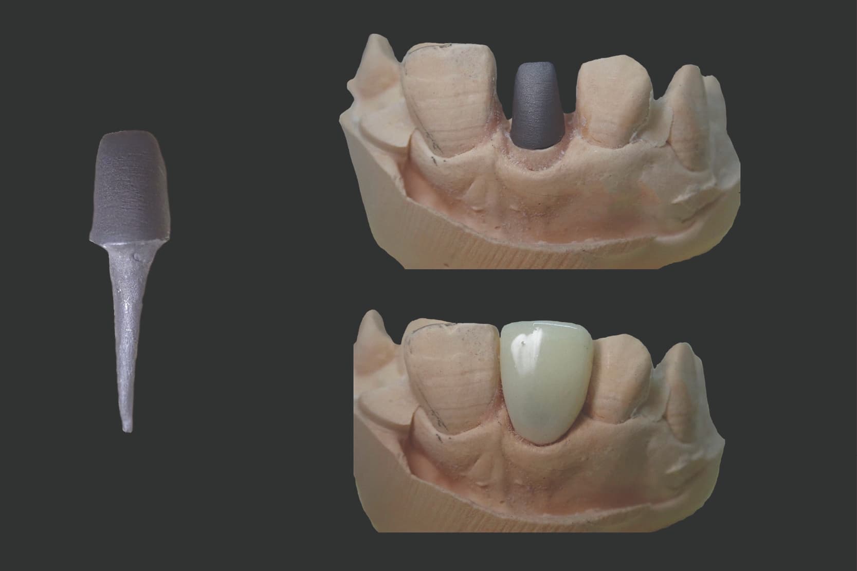
Endodontic posts can be either prefabricated or custom made.
Custom-made or cast dental posts have been considered the gold standard for the rehabilitation of devitalised teeth with severe coronal structure loss for some years now. Custom-made dental posts have a shape that is more similar to the actual anatomy of the root canal and provide optimum retention and support for the coronal prosthesis. (2) Dental posts are therefore used to restore devitalised teeth (3) and provide mechanical support for the subsequent prosthetic restoration.
Although fibreglass (prefabricated) posts have shown comparable long-term survival to that of cast (custom made) posts (4-6), custom-made dental posts are often chosen in cases of severe coronal tissue loss when the splint is less than 2 mm or when the element is a rehabilitation pillar with long spans. (7)
Although cast dental posts have a higher failure strength than fibreglass posts (8), at the same time they express very different elastic modulus values to those of the dentin tissue they are connected to and this difference in elasticity can contribute to the unbonding of this type of posts and their higher root fracture risk. (9)
How are custom-made dental posts fabricated?
There are traditionally two techniques: indirect and direct. (10)
Indirect technique
Step 1 – Having established that the root canal therapy and root canal filling have been carried out properly, use special burs prepare the space for the post by removing root canal filling material to obtain the right diameter and depth.
Step 2 – Once preparation is complete, proceed with the actual impression taking phase, which usually consists of a one-step technique. Using a thin tip, inject low-density elastomeric material into the endodontic space and onto the preparation; then position an impression tray previously loaded with a high-density impression material, and take the impression.
Step 3 – The dental technician makes the gypsum model, on which they make the dental post, using a wax-up. They then use a lost-wax casting technique to fabricate the metal dental post.
These are several variants of this technique. In some cases, the low-density elastomeric material is dispensed into the root canal using a lentulo spiral, or a castable post is used as a physical aid with which to introduce the material deeper into the root canal.
Direct technique
Following completion of the preparation, which must have the characteristics described above, it is possible to proceed with the fabrication phase.
The direct technique does not include an impression phase as such.
Step 1 – Choose a suitable sized castable post that can be introduced deep into the canal without causing tension, and allows passive introduction and passage, as well as repeatable positioning.
Step 2 – Once the post has been chosen and modified if necessary, prepare the acrylic resin. The density of the resin must be such that it can be inserted into the root canal space and, at the same time allow a certain degree of mouldability. Sink the castable post into the resin and position it inside the root canal then, using a small spatula, shape the coronal part to form the abutment.
Step 3 – Wait for the resin to set, remove the dental post and repeat the procedure in order to reproduce the anatomy of the root canal more accurately. Once this step is complete, use a diamond bur to prepare and smoothen the coronal surface of the abutment.
Step 4 – Lastly, send the fabricated piece to the dental technician, who will complete the process by fabricating a metal duplicate.
Which technique gives the better results?
Although one in vitro study showed that there are no significant differences in terms of accuracy, the indirect technique reduces chair time. (10)
According to another study, both the direct technique and the indirect technique result in metal dental posts that are undersized but nevertheless suitable for bonding. According to the same study, the indirect technique affords shorter chair times and is therefore to be preferred when multiple rehabilitations are required. (11)
How do optical impressions perform for this application?
A recent in vitro study showed that, at the current time, optical impressions have many limits resulting from the depth and very limited diameter of the root canal.
As a matter of fact, with this type of application, the scanner light is unable to penetrate into the most apical part, or reach the narrow walls of the post space, meaning that conventional impressions are almost always necessary. (12)
The deeper and narrower the root canal is, the more difficult it will be for the light to reach all the surfaces and, therefore, to read the anatomy correctly.
Recently, a hybrid method consisting of a conventional impression of the endodontic space and its subsequent scanning was proposed in order to fabricate custom-made dental posts using digital CAD/CAM techniques. (13)
Conclusions
Very often, dental posts are the last feasible option for rehabilitating a severely compromised tooth.
Although there are various alternatives, this technique is still very much in favour and it constitutes a safe method that is validated by years of literature. There are two main fabrication techniques, but it is possible to find several variants of both.
At the current time, the digital technique, particularly in certain cases, would not appear able to deliver the same levels of accuracy as the conventional technique, due to the unfavourable conditions resulting from the anatomy of the root canal space.
References
- Al-Omiri MK, Mahmoud AA, Rayyan MR, Abu-Hammad O. Fracture resistance of teeth restored with post-retained restorations: an overview. J Endod 2010;36:1439-49.
- Ravanshad S, Ghoreeshi N. An in vitro study of coronal microleakage in endodontically-treated teeth restored with posts. Aust Endod J 2003;29: 128-33.
- Durmus G, Oyar P. Effects of post core materials on stress distribution in the restoration of mandibular second premolars: a finite element analysis. J Prosthet Dent 2014;112:547-54
- Sarkis-Onofre R, Jacinto Rde C, Boscato N, Cenci MS, Pereira-Cenci T. Cast metal vs. glass fibre posts: a randomized controlled trial with up to 3 years of follow up. J Dent 2014;42:582-7.
- Cormier CJ, Burns DR, Moon P. In vitro comparison of the fracture resistance and failure mode of fiber, ceramic, and conventional post systems at various stages of restoration. J Prosthodont 2001;10:26-36.
- Hu H, Pang LC, Hsu CC, Lau YH. Fracture resistance of endodontically treated anterior teeth restored with four post-and-core systems. Quintessence Int 2003;34:349-53.
- Heydecke G, Peters CM. The restoration of endodontically treated, singlerooted teeth with cast or direct posts and cores: a systematic review. J Prosthet Dent 2002;87:380-6.
- Zhou L, Wang Q. Comparison of Fracture Resistance between Cast Posts and Fiber Posts: A Meta-analysis of Literature. J Endod, 2013;39:11-15.
- Ona M, Wakabayashi N, Yamazaki T, Takaichi A, Igarashi Y. The influence of elastic modulus mismatch between tooth and post and core restorations on root fracture. Int Endod J., 2013 Jan;46:47-52.
- Rayyan, M. R., Roa’a, A. A., Alsadun, S. F., & Hijazy, F. R. (2016). Accuracy of cast posts fabricated by the direct and the indirect techniques. The Journal of prosthetic dentistry, 116(3), 411-415.
- de Moraes, A. P., Neto, V. P., Boscato, N., & Pereira-Cenci, T. (2016). Randomized clinical trial of the influence of impression technique on the fabrication of cast metal posts. The Journal of Prosthetic Dentistry, 116(1), 47-51.
- Pinto, A., Arcuri, L., Carosi, P., Nardi, R., Libonati, A., Ottria, L., & Campanella, V. (2017). In vitro evaluation of the post-space depth reading with an intraoral scanner (IOS) compared to a traditional silicon impression. Oral & Implantology, 10(4), 360.
- Spina, D. R. F., da Costa, R. G., Correr, G. M., & Rached, R. N. (2018). Scanning of root canal impression for the fabrication of a resin CAD-CAM-customized post-and-core. The Journal of prosthetic dentistry, 120(2), 242-245.
Do you want more information on Zhermack Dental products and solutions?
Contact us


