In recent years there has been an increase in the interest in digital technologies that has impacted various sectors worldwide, from defence to engineering, and even the health sector, including dentistry.
More specifically, the advent of CAD-CAM (computer-aided design/computer-assisted manufacturing) systems has led to the development of digital technologies for intraoral impression-taking. This process involves the use of a scanner that acts as a tool for acquiring data in order to produce three-dimensional (3D) images of the structures (teeth, dental arches and tissues) scanned, in the same way as with an analogical impression.
The digital impression systems now used in daily clinical practice afford higher performance in terms of accuracy and precision, although in some cases analogical impressions remain irreplaceable and decisive for giving the technician the correct clinical information.
The advantages of digital impressions
Optical impression-taking using an intraoral scanner (IOS) presents a series of advantages and resolves certain issues that characterise conventional impression-taking1. As a matter of fact, digital impressions allow, for example, faster and, in some cases, better communication with dental technicians and patients2.
Digital impressions do away with the gypsum model-making step, which involves possible errors in analogical impression casting and the impression can be kept as an STL file for an unlimited period of time. It also affords extremely direct access to the digital work-flow, and to CAD/CAM systems with subtractive and additive technologies, which are now always used by dental technicians to materially produce prostheses and mesostructures.
Digital impressions also avoid the discomfort that impression materials can cause patients, such as bouts of vomiting and the unpleasant taste, simultaneously also eliminating the risk of presenting allergies to the impression material3,4. On a technical level, digital impressions are taken more quickly by dentists, especially when compared with impressions taken using manually-mixed materials, although the result is very operator-dependent.

The advantages of analogical impressions
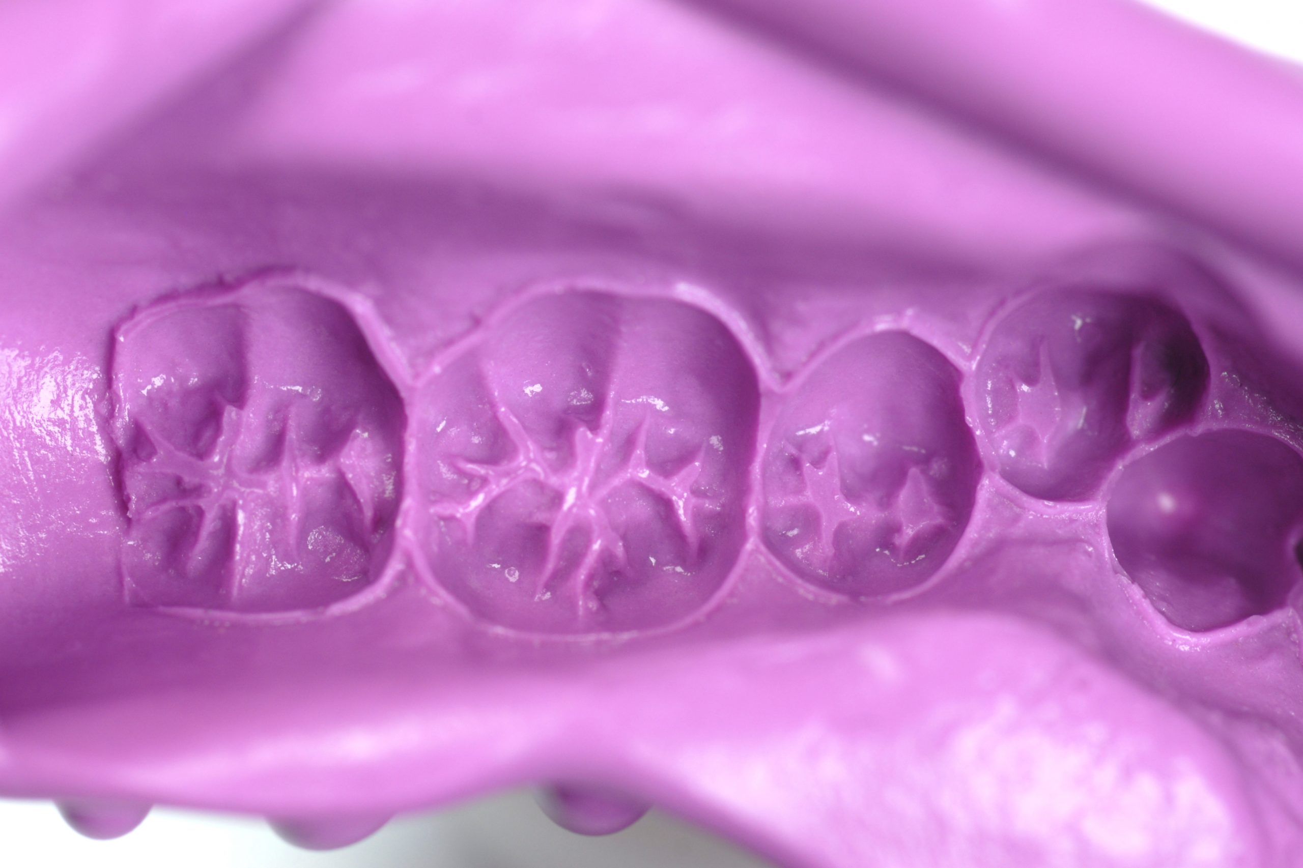
Analogical impressions are still the impression type most commonly used in the majority of dental practices5. The reasons for this lie, first and foremost, in the fact that the cost of analogical impressions is far lower than the cost of digital impressions (regardless of whether the impression is taken using materials like alginate or whether elastomers are used).
Intraoral scanners are extremely expensive, not only at the time of purchase, but also afterwards, as they often entail annual costs for upgrades and assistance. This means that dentists need to obtain important returns on the investment, which they might not be able to guarantee in the short term.
Then there are aspects regarding digitalisation, which involves a sometimes steep learning curve6, especially for those dentists who are less familiar with new technologies and still account for a significant part of the dentistry sector.
Furthermore, despite the great accuracy and precision that various scanners available on the market now afford, in certain clinical situations impression materials are still the first choice7.
Digital and analogical impressions compared
An interesting in vivo study comparing intraoral scanners and conventional impressions using alginate in edentulous maxillae7, showed that analogical impression-taking is more accurate than digital impressions in capturing the peripheral soft tissues in order to establish the marginal peripheral seal areas of removable prostheses.
The authors therefore concluded that intraoral scanners may thus only be considered valid for replacing the preliminary impression, in order to obtain a model on which to build an individual impression. In view of permanent mobile prosthetic restoration, on the other hand, it is essential to exert pressure in the areas of the peripheral soft tissues, which is currently impossible without a mucodynamic impression.
Another clinical situation in which digital systems are less appropriate than conventional impressions is when the end of preparation margins to be indicated on the teeth are subgingival or iuxtagingival8 or in cases in which the gingival margin bleeds.
Implant prostheses, on the other hand, represent a separate and highly controversial chapter. In these cases, especially when it is necessary to perform rehabilitation of the whole arch on multiple implants, a number of studies agree that not all intraoral scanners are suited to this purpose9. Furthermore, the larger the arch, the heavier the output STL file that the scanner will have to export (and scanners have exportation limits).
In any case, when it comes to impressions on multiple implants, the clinical detection time of an optical impression is far shorter than that required for analogical impression-taking10. It is also important not to overlook the potential difficulties in removing analogical impressions from the patient’s mouth in cases in which the implants are not parallel to one another, difficulties that can cause distortions in the impression material around the transfers3,11.
This is not the case for fixed prostheses on natural teeth where conventional whole-arch impressions are far less demanding in terms of time and preferred over intraoral scans by both dentists and patients12.
Conclusions
Digital impressions have come to be a useful and advantageous work tool in daily clinical practice. Many of the defects that can be encountered with impression materials are now overcome by digital impressions with undeniable clinical and temporal advantages. However, analogical impression-taking is still considered the gold standard in certain clinical situations and the more commonly used technique in dentistry.
In the future it will be interesting to understand how to overcome the defects of both methods by integrating the two impression-taking techniques.
References
1. Zimmermann, M., Mehl, A., Mörmann, W. H. & Reich, S. Intraoral scanning systems – a current overview. Int. J. Comput. Dent. 18, 101–129 (2015).
2. Goracci, C., Franchi, L., Vichi, A. & Ferrari, M. Accuracy, reliability, and efficiency of intraoral scanners for full-arch impressions: a systematic review of the clinical evidence. Eur. J. Orthod. 38, 422–428 (2016).
3. Yuzbasioglu, E., Kurt, H., Turunc, R. & Bilir, H. Comparison of digital and conventional impression techniques: evaluation of patients’ perception, treatment comfort, effectiveness and clinical outcomes. BMC Oral Health 14, 10 (2014).
4. Wismeijer, D., Mans, R., van Genuchten, M. & Reijers, H. A. Patients’ preferences when comparing analogue implant impressions using a polyether impression material versus digital impressions (Intraoral Scan) of dental implants. Clin. Oral Implants Res. 25, 1113–1118 (2014).
5. Cervino, G. et al. Alginate Materials and Dental Impression Technique: A Current State of the Art and Application to Dental Practice. Mar. Drugs 17, (2018).
6. Mangano, F., Gandolfi, A., Luongo, G. & Logozzo, S. Intraoral scanners in dentistry: a review of the current literature. BMC Oral Health 17, 149 (2017).
7. D’Arienzo, L. F., D’Arienzo, A. & Borracchini, A. Comparison of the suitability of intra-oral scanning with conventional impression of edentulous maxilla in vivo. A preliminary study. J. Osseointegration 10, 115–120 (2018).
8. Mangano, F. G., Margiani, B., Solop, I., Latuta, N. & Admakin, O. An Experimental Strategy for Capturing the Margins of Prepared Single Teeth with an Intraoral Scanner: A Prospective Clinical Study on 30 Patients. Int. J. Environ. Res. Public. Health 17, (2020).
9. Di Fiore, A. et al. Full arch digital scanning systems performances for implant-supported fixed dental prostheses: a comparative study of 8 intraoral scanners. J. Prosthodont. Res. 63, 396–403 (2019).
10. Lee, S. J. & Gallucci, G. O. Digital vs. conventional implant impressions: efficiency outcomes. Clin. Oral Implants Res. 24, 111–115 (2013).
11. Baig, M. R. Multi-unit implant impression accuracy: A review of the literature. Quintessence Int. Berl. Ger. 1985 45, 39–51 (2014).
12. Sailer, I., Mühlemann, S., Fehmer, V., Hämmerle, C. H. F. & Benic, G. I. Randomized controlled clinical trial of digital and conventional workflows for the fabrication of zirconia-ceramic fixed partial dentures. Part I: Time efficiency of complete-arch digital scans versus conventional impressions. J. Prosthet. Dent. 121, 69–75 (2019).
Do you want more information on Zhermack Dental products and solutions?
Contattaci


 Zhermack SpA has been one of the most important producers and international distributors of alginates, gypsums and silicone compounds for the dental sector for over 40 years. It has also developed solutions for the industrial and wellbeing sectors.
Zhermack SpA - Via Bovazecchino, 100 - 45021 Badia Polesine (RO), Italy.


