Implant-supported fixed prostheses include all those dental prostheses such as crowns, full-arch prostheses and partial prostheses, as well as maxillofacial prostheses, that are anchored to and entirely supported by dental implants [1].
Clinical and technical phases of the fabrication of multiple-implant prostheses
The clinical and technical techniques for making multiple-implant prostheses consist of a series of rehabilitation steps that, depending on the type of prosthesis, materials and techniques used, require good knowledge of the subject-matter [2–4].
First of all, it should be pointed out that the clinical phases for making multiple-implant prostheses, be they cement- or screw-retained, can vary depending on the clinical case and the type of prosthetic restoration [5].
This article focuses solely on the conventional clinical procedures carried out for multiple-implant restorations and does not cover immediate loading or provisional restorations.
Conventional clinical procedures
The conventional clinical procedures can be summarised as:
- implant uncovering;
- impression;
- testing of the structure connecting the implants;
- testing of the final restoration;
- delivery.
We will now take a more detailed look at each one.
Implant uncovering phase
The first phase is implant uncovering (for submerged implants).
This step, which is carried out once the osteointegration resulting from implant placement is complete, consists in surgically uncovering the implant emergences, removing the cover screws protecting the connections and subsequently placing the healing screws on the implants.
This phase is by-passed when implants with a smooth transmucosal collar (tissue-level implants) are inserted, as the adaptation of the mucosae to the implant collar takes place at the same time as osteointegration [6].
Following the surgical uncovering of the implant emergences, it is necessary to wait for the soft tissues to readjust and heal properly around the healing screws.
The time required varies from one individual to another, but also depend on the type of uncovering technique used, the surgical manoeuvres, the type of healing screws applied and the amount of keratinised tissue [7].
This is necessary and fundamental for the subsequent implant impression phase.
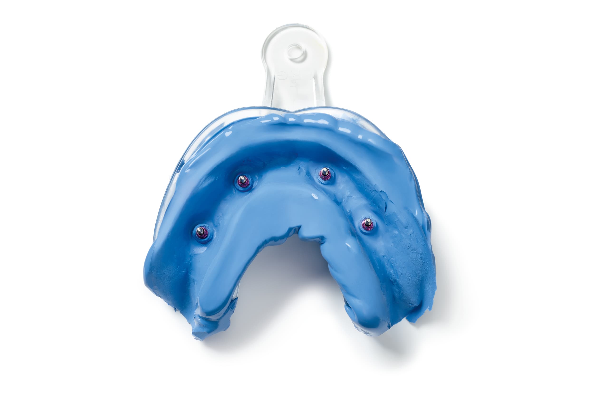
Implant impression phase
In the implant impression phase, in addition to the spatial three-dimensional position of the connections, it is also necessary to capture the profiles and the positions of the adjacent peri-implant and inter-implant soft tissues.
However, the real challenge of implant impressions regards the proper three-dimensional reproduction of the position of the connections in the technician’s work model [8–10].
Testing of the structure connecting the implants
An accurate reproduction of the positions of the connections in the 3D model or gypsum model is the first step to fabricating a “passive” prosthesis, i.e. a prosthesis that is free from tensions once it is screwed into the implants or intermediate abutments [11].
Prosthetic imprecision is undeniably dangerous, as it increases the risk of mechanical complications such as loss of connection screw torque, connection screw fracture, and the chipping or fracture of the coating materials and/or the prosthetic components [12–14].
It is not clear whether a lack of passivity also poses a risk for the implants; however, checking for the absence of tensions when screwing in the framework is a must for multi-implant restorations [15].
In this regard, impressions on implant-supported prostheses, especially for full-arch prostheses, appear to be taken with polyethers or polyvinylsiloxanes and with the open-tray pick-up technique in most cases [8,16].
Want to find out more on this topic? Read the post on pick-up impressions and closed-tray impressions.
Even if the impression is taken properly, the laboratory procedures for fabricating a prosthesis connecting multiple implants can in turn introduce errors, depending on the materials and the framework production techniques used [17].
The passivity of the structure connecting the implants must therefore always be tested and clinically assessed [18].
This is usually done using the “Sheffield Test”, which consists in screwing the structure to the most distal or mesial implant, before clinically and radiographically checking the passivity of the framework on the contralateral implant [12].
Once this step has been carried out, it is possible to proceed with the subsequent prosthetic finalisation and fit test steps, following the application of the coating material [17].
Testing of the final restoration
In this step, it is necessary to check both the interdental contact points and occlusion.
As regards the latter, in prosthodontics there are different schools of thought and solutions, which are proposed depending on the individual scenarios [19,20]. It is, however, important to point out that the absence of periodontal receptors makes dental implants far more susceptible to occlusal overloadingthan natural teeth [19,21].
Therefore, given the inability to absorb the masticatory load and the impossibility of fine occlusal force adaptation, it becomes particularly important to control the occlusion of full-arch implant-supported prostheses [19–21].
In this regard, a balanced occlusion with simultaneous bilateral contacts of equal intensity on the posterior elements, maintenance of clearance (10 µm) on the distal cantilevers and on the anterior teeth, the elimination of precontacts and interferences, and control of the release guides and “centric freedom” constitute a fundamental prerequisite for a satisfactory full-arch restoration also at occlusal level [19–21].
Delivery of the final restoration
Once this clinical phase has been managed, it is possible to proceed with the delivery of the final restoration, which will nevertheless always require future maintenance, check-ups and maintenance hygiene intervention, both at home and, above all, by a professional, in order to enhance the durability of the restoration.
High-performance solutions for impression-taking
Zhermack provides high-performance solutions for impression-taking on both implants and natural stumps. On the one hand, Hydrorise Implant is scannable and affords ideal rigidity for impression-taking on implants. On the other, Hydroriseguarantees fine detail reproduction and excellent hydrocompatibility, which contribute to the precision and accuracy of natural stump impressions.
References
[1] The Glossary of Prosthodontic Terms: Ninth Edition. J Prosthet Dent 2017;117: e1-e105 n.d.
[2] Ortensi L, Ortensi M, Minghelli A, Grande F. Implant-Supported Prosthetic Therapy of an Edentulous Patient: Clinical and Technical Aspects. Prosthesis 2020;2:140–52. https://doi.org/10.3390/prosthesis2030013.
[3] Montanari M, Grande F, Lepidi L, Piana G, Catapano S. Rehabilitation with implant-supported overdentures in preteens patients with ectodermal dysplasia: A cohort study. Clinical Implant Dentistry and Related Research n.d.;n/a. https://doi.org/10.1111/cid.13258.
[4] Catapano S, Ferrari M, Mobilio N, Montanari M, Corsalini M, Grande F. Comparative Analysis of the Stability of Prosthetic Screws under Cyclic Loading in Implant Prosthodontics: An In Vitro Study. Applied Sciences 2021;11:622. https://doi.org/10.3390/app11020622.
[5] Lops, D., Bruna, E., & Fabianelli, A. (2014). La protesi implantare. Dental Cadmos, 6(82), 386. n.d.
[6] Fernández-Formoso N, Rilo B, Mora MJ, Martínez-Silva I, Díaz-Afonso AM. Radiographic evaluation of marginal bone maintenance around tissue level implant and bone level implant: a randomised controlled trial. A 1-year follow-up. Journal of Oral Rehabilitation 2012;39:830–7. https://doi.org/10.1111/j.1365-2842.2012.02343.x.
[7] Mattheos N, Vergoullis I, Janda M, Miseli A. The Implant Supracrestal Complex and Its Significance for Long-Term Successful Clinical Outcomes. Int J Prosthodont 2021;34:88–100. https://doi.org/10.11607/ijp.7201.
[8] Baldissara P, Koci B, Messias AM, Meneghello R, Ghelli F, Gatto MR, et al. Assessment of impression material accuracy in complete-arch restorations on four implants. J Prosthet Dent 2021. https://doi.org/10.1016/j.prosdent.2020.10.017.
[9] Tallarico M, Galiffi D, Scrascia R, Gualandri M, Zadrożny Ł, Czajkowska M, et al. Digital Workflow for Prosthetically Driven Implants Placement and Digital Cross Mounting: A Retrospective Case Series. Prosthesis 2022;4:353–68. https://doi.org/10.3390/prosthesis4030029.
[10] Alsharbaty MHM, Alikhasi M, Zarrati S, Shamshiri AR. A Clinical Comparative Study of 3‐Dimensional Accuracy between Digital and Conventional Implant Impression Techniques. Journal of Prosthodontics 2019;28:e902–8. https://doi.org/10.1111/jopr.12764.
[11] Grande F, Cesare PM, Zamperoli EM, Gianoli CM, Mollica F, Catapano S. Evaluation of Tension and Deformation in a Mandibular Toronto Bridge Anchored on Three Fixtures Using Different Framework Materials, Abutment Systems, and Loading Conditions: A FEM Analysis. Eur J Dent 2023. https://doi.org/10.1055/s-0042-1758785.
[12] Rutkunas V, Dirse J, Kules D, Mischitz I, Larsson C, Janda M. Misfit simulation on implant prostheses with different combinations of engaging and nonengaging titanium bases. Part 2: Screw resistance test. J Prosthet Dent 2022:S0022-3913(22)00286-4. https://doi.org/10.1016/j.prosdent.2022.04.027.
[13] Ortorp A, Jemt T, Wennerberg A, Berggren C, Brycke M. Screw preloads and measurements of surface roughness in screw joints: an in vitro study on implant frameworks. Clin Implant Dent Relat Res 2005;7:141–9. https://doi.org/10.1111/j.1708-8208.2005.tb00058.x.
[14] Jemt T. A retro-prospective effectiveness study on 3448 implant operations at one referral clinic: A multifactorial analysis. Part II: Clinical factors associated to peri-implantitis surgery and late implant failures. Clin Implant Dent Relat Res 2017;19:972–9. https://doi.org/10.1111/cid.12538.
[15] Goodacre CJ, Bernal G, Rungcharassaeng K, Kan JYK. Clinical complications with implants and implant prostheses. J Prosthet Dent 2003;90:121–32. https://doi.org/10.1016/S0022-3913(03)00212-9.
[16] Schmidt A, Häussling T, Rehmann P, Schaaf H, Wöstmann B. Accuracy of various impression materials and methods for two implant systems: An effect size study. J Prosthodont Res 2018;62:245–51. https://doi.org/10.1016/j.jpor.2017.10.004.
[17] Revilla-León M, Sánchez-Rubio JL, Pérez-López J, Rubenstein J, Özcan M. Discrepancy at the implant abutment-prosthesis interface of complete-arch cobalt-chromium implant frameworks fabricated by additive and subtractive technologies before and after ceramic veneering. J Prosthet Dent 2021;125:795–803. https://doi.org/10.1016/j.prosdent.2020.03.018.
[18] Darós P, Carneiro VC, Siqueira AP, de-Azevedo-Vaz SL. Diagnostic accuracy of 4 intraoral radiographic techniques for misfit detection at the implant abutment joint. The Journal of Prosthetic Dentistry 2018;120:57–64. https://doi.org/10.1016/j.prosdent.2017.08.008.
[19] Kim Y, Oh T-J, Misch CE, Wang H-L. Occlusal considerations in implant therapy: clinical guidelines with biomechanical rationale. Clin Oral Implants Res 2005;16:26–35. https://doi.org/10.1111/j.1600-0501.2004.01067.x.
[20] Yoon D, Pannu D, Hunt M, Londono J. Occlusal considerations for full-arch implant-supported prostheses: A guideline. Dentistry Review 2022;2:100042. https://doi.org/10.1016/j.dentre.2022.100042.
[21] Hämmerle CH, Wagner D, Brägger U, Lussi A, Karayiannis A, Joss A, et al. Threshold of tactile sensitivity perceived with dental endosseous implants and natural teeth. Clin Oral Implants Res 1995;6:83–90. https://doi.org/10.1034/j.1600-0501.1995.060203.x.
Do you want more information on Zhermack Dental products and solutions?
Contact us


