12
Jun
Analogue and digital workflows for removable full dentures: the main differences
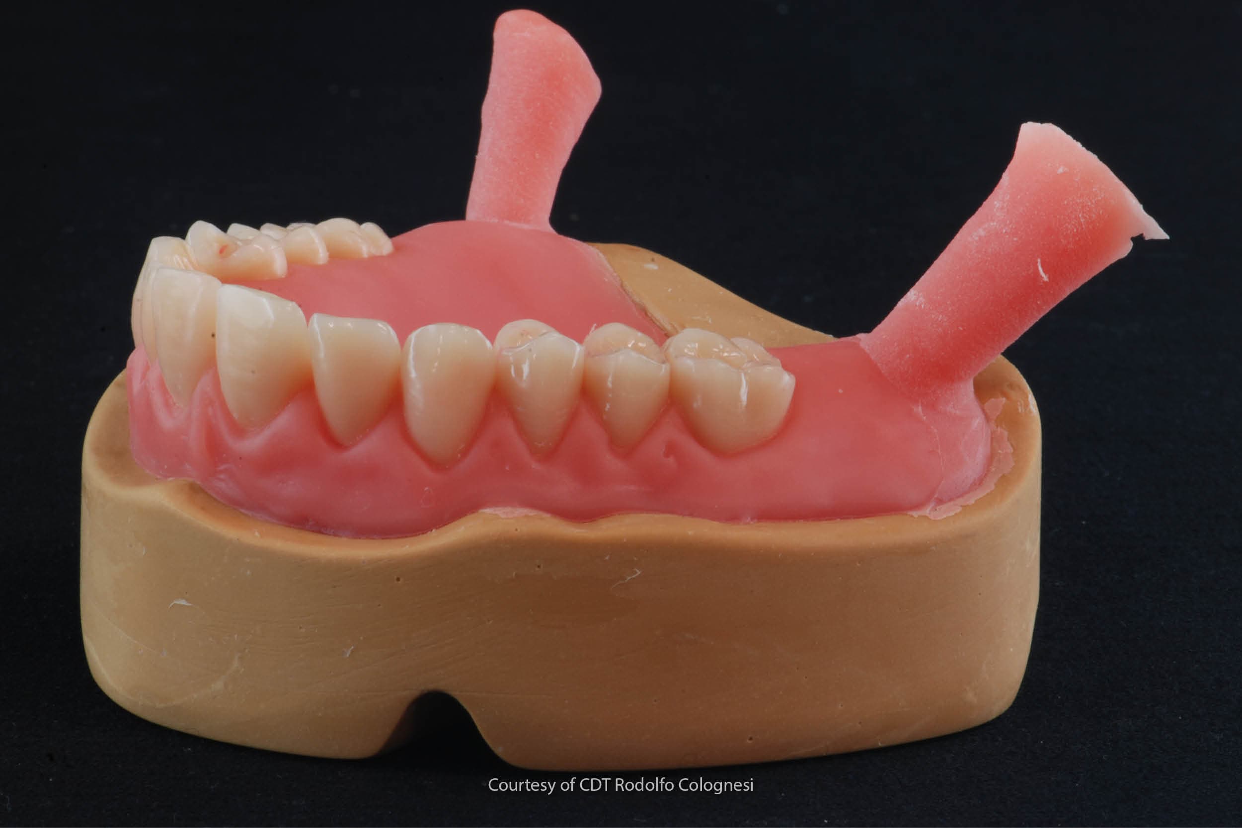
Removable full dentures are full dental prostheses that replace the entire dentition and the reabsorbed soft and hard tissues of the upper and lower jaws, and that can be easily inserted and removed by patients themselves [1].
The clinical and technical stages required to fabricate removable full dentures are a great challenge for dentists and dental technicians, as the many steps and the handling of different materials and techniques require of both of them excellent manual skills combined with considerable theoretical knowledge [2,3].
The analogue clinical and laboratory prosthetic workflow remains the gold standard for the rehabilitation with a removable full denture of a patient in whom one or both arches are edentulous [4,5].
The digital workflow only has advantages over the analogue workflow in some of the steps of these rehabilitations. Let’s take a closer look at them.
The advantages of the digital workflow over the “gold standard” analogue workflow in certain stages of rehabilitation
The rehabilitation steps in which the digital workflow has advantages over the analogue one are primarily technical steps, which digitally simplify the dental technician’s handling of the materials and improve the biocompatibility and chemical and physical characteristics of the resins used to fabricate the prostheses [6–8].
It has been demonstrated that the tissue congruency of CAD-CAM milled removable full dentures is superior to that of prostheses fabricated using the conventional flasking technique, in that milling a pre-cured disk of PMMA prevents the curing-induced shrinkage typical of acrylic resins resulting from the transition from cold to hot [3,8–10].
The 3D printing of denture bases 3D printing of denture bases also appears to give promising results in terms of their congruence with the denture-bearing tissues, although the difference between the various printers and the different types of resin do not make it possible to confirm its superiority over the conventional techniques for the fabrication of removable dentures [8].
The 9 steps of the conventional technique
There are currently a great many analogue workflows for removable full dentures. For the sake of simplicity, however, we can consider these workflows to be variants of the conventional linear technique [11].
This technique, as has already been extensively described [11], consists of a total of 9 steps (5 clinical and 4 technical steps):
- the edentulous patient’s first appointment and taking of preliminary alginate impressions of the arches;
- development of the preliminary gypsum models and fabrication of the custom impression trays;
- functional secondary impressions;
- development of the master models and fabrication of the occlusion rims;
- recording of the centric relation;
- mounting of the models with the occlusion rims on an articulator, choice and assembly of the frontal elements and diatorics;
- aesthetic, phonetic and functional testing and patient consent;
- finishing of the dentures in the laboratory;
- delivery of the denture to the patient following a fit check.
This workflow cannot be completely translated into a digital workflow due to the limitations of intraoral scanners (already partly discussed in the article “Digital impressions vs analogical impressions: when to use one and when the other”) [12,13].
The analogue steps for the fabrication of functional secondary impressions and master models for the fabrication of wax occlusion rims remain a fundamentally analogue step that still cannot be replaced by digital techniques.
Lab Procedure for complete removable prosthesis with hot-curing resins:
The possible digital workflows that can be used for removable full dentures
The possible digital workflows that can be used for removable full dentures do not contemplate a “full-digital workflow”, rather only mixed analogue-digital workflows that can be classified into 4 categories [14]:
- Analogue-digital workflows with a predominant analogue component. In this workflow, all the steps are carried out analogically, but the final denture is fabricated by milling based on a CAD file obtained by scanning resin plates in which the teeth are fitted with the proper clinically-detected vertical dimension.
- If, on the other hand, the patient already has dated removable dentures that have become incongruent, they can be used to re-establish the proper centric relation and to obtain a new impression of the soft tissues. The whole block can then be scanned by the dental technician in order to switch to a digital workflow and fabricate, again using a CAD-CAM technique, the final dentures.
- If, on the hand, multiple extractions are required in patients who do not want to remain toothless, the digital approach can be useful for rapidly fabricating pre-extraction full dentures by 3D printing based on an intraoral scan or a subsequently-scanned alginate impression. The same prosthesis can then be relined inside the patient’s mouth with normal resin materials.
- Analogue-digital workflows with a predominant digital component. This flow starts with an analogue alginate impression that is used to obtain models that are subsequently scanned. Alternatively, it is possible to scan the impression itself in order to fabricate a 3D-printed try-in. This device is applied to the oral cavity to record the centric relation, in order to identify the gothic arch with the mandibular movements on the horizontal plane and to take functional impressions using an impression material. The digital data are then imported by scanning the whole block and the final removable dentures are fabricated using a CAD-CAM technique.
Alginates for removable dentures in edentulous patients
Although these workflows can constitute a new beginning for treatment with removable prosthetics, they nevertheless always require a dental laboratory that is equipped for digital workflows, i.e. with the appropriate modules in the various software programmes.
It is also essential for the dental technician to have an in-depth knowledge of the digital workflow and the errors associated with integrating analogue workflows into CAD-CAM systems. For this reason, further research is required in order to obtain a more comprehensive understanding of the indications and limitations of new technologies in this type of rehabilitation.
Zhermack manufactures a vast range of alginates with characteristics that are suited to use in partly or fully edentulous patients for the fabrication of partial and full removable dentures.
More specifically, Neocolloid is an alginate with a long setting time designed for obtaining adequate impressions of the mucous membranes of edentulous patients and that, thanks to its physical and chemical characteristics, allows an excellent reproduction of the maxillary mucosae.
Bibliography
[1] The Glossary of Prosthodontic Terms: Ninth Edition. J Prosthet Dent 2017;117: e1-e105 n.d.
[2] Ortensi L, Ortensi M, Minghelli A, Grande F. Implant-Supported Prosthetic Therapy of an Edentulous Patient: Clinical and Technical Aspects. Prosthesis 2020;2:140–52. https://doi.org/10.3390/prosthesis2030013.
[3] Grande F, Tesini F, Pozzan MC, Zamperoli EM, Carossa M, Catapano S. Comparison of the Accuracy between Denture Bases Produced by Subtractive and Additive Manufacturing Methods: A Pilot Study. Prosthesis 2022;4:151–9. https://doi.org/10.3390/prosthesis4020015.
[4] Srinivasan M, Kalberer N, Fankhauser N, Naharro M, Maniewicz S, Müller F. CAD-CAM complete removable dental prostheses: A double-blind, randomized, crossover clinical trial evaluating milled and 3D-printed dentures. J Dent 2021;115:103842. https://doi.org/10.1016/j.jdent.2021.103842.
[5] Peroz S, Peroz I, Beuer F, Sterzenbach G, von Stein-Lausnitz M. Digital versus conventional complete dentures: A randomized, controlled, blinded study. J Prosthet Dent 2022;128:956–63. https://doi.org/10.1016/j.prosdent.2021.02.004.
[6] Wagner SA, Kreyer R. Digitally Fabricated Removable Complete Denture Clinical Workflows using Additive Manufacturing Techniques. Journal of Prosthodontics 2021;30:133–8. https://doi.org/10.1111/jopr.13318.
[7] Goodacre CJ, Goodacre BJ, Baba NZ. Should Digital Complete Dentures Be Part of A Contemporary Prosthodontic Education? J Prosthodont 2021;30:163–9. https://doi.org/10.1111/jopr.13289.
[8] Hwang H-J, Lee SJ, Park E-J, Yoon H-I. Assessment of the trueness and tissue surface adaptation of CAD-CAM maxillary denture bases manufactured using digital light processing. J Prosthet Dent 2019;121:110–7. https://doi.org/10.1016/j.prosdent.2018.02.018.
[9] Faty MA, Sabet ME, Thabet YG. A comparison of denture base retention and adaptation between CAD-CAM and conventional fabrication techniques. Int J Prosthodont 2022. https://doi.org/10.11607/ijp.7193.
[10] Goodacre BJ, Goodacre CJ, Baba NZ, Kattadiyil MT. Comparison of denture base adaptation between CAD-CAM and conventional fabrication techniques. J Prosthet Dent 2016;116:249–56. https://doi.org/10.1016/j.prosdent.2016.02.017.
[11] Moderno Trattato di protesi Mobile Completa [Glauco – Martina Edizioni] n.d. https://www.medicalinformation.it/moderno-trattato-di-protesi-mobile-completa-glauco-martina-edizioni-9788875721183glauco-marino-canton-alessandro-marino-antonino-di-lullo-nicola.html (accessed January 3, 2023).
[12] D’Arienzo LF, D’Arienzo A, Borracchini A. Comparison of the suitability of intra-oral scanning with conventional impression of edentulous maxilla in vivo. A preliminary study. Journal of Osseointegration 2018;10:115–20. https://doi.org/10.23805/jo.2018.10.04.02.
[13] Mangano F, Gandolfi A, Luongo G, Logozzo S. Intraoral scanners in dentistry: a review of the current literature. BMC Oral Health 2017;17:149. https://doi.org/10.1186/s12903-017-0442-x.
[14] Maragliano-Muniz P, Kukucka ED. Incorporating Digital Dentures into Clinical Practice: Flexible Workflows and Improved Clinical Outcomes. J Prosthodont 2021;30:125–32. https://doi.org/10.1111/jopr.13277.
Do you want more information on Zhermack Dental products and solutions?
Contact us

 Zhermack SpA has been one of the most important producers and international distributors of alginates, gypsums and silicone compounds for the dental sector for over 40 years. It has also developed solutions for the industrial and wellbeing sectors.
Zhermack SpA - Via Bovazecchino, 100 - 45021 Badia Polesine (RO), Italy.


