Atrophy of the alveolar process following tooth loss leads, in some patients, to the replacement of bone tissue with hyperplastic fibrous tissue, known as ‘flabby tissue’. (1)
This condition is most frequently observed in the maxilla, especially in the anterior region, with a reported prevalence of 24%, but it may also occur in the mandibular area, where the prevalence is 5%. (2,3)
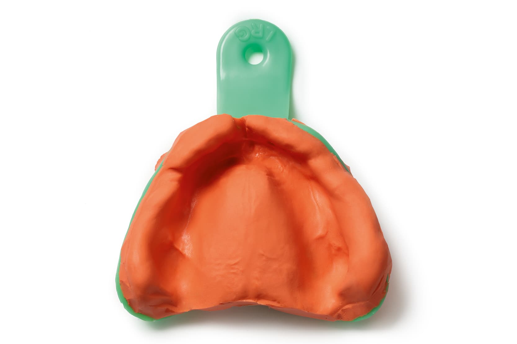
Floating ridges and the challenges of recording soft tissues
Accurate recording of edentulous soft tissues is a fundamental and necessary step in achieving a stable, retentive, and comfortable complete denture. (4) In this respect, the presence of flabby ridges or excessively mobile tissues over edentulous bony structures represents one of the main challenges for the clinician providing complete dentures, since these areas tend to deform under the pressure exerted during impression taking, altering their true morphology and compromising the stability of the future denture. (5)
Flabby tissue also tends to move under functional load, causing micromovements of the denture base, loss of stability, and the formation of traumatic lesions. It is therefore essential to record the tissue in its physiological position, avoiding compressions that would alter its shape and compromise the congruity of the denture-base fit.
Impression techniques in the edentulous patient
Over the years, several impression philosophies have been proposed for edentulous patients: mucostatic, mucocompressive, and selective-pressure. (5)
- The mucostatic technique aims to record tissues at rest, exploiting surface tension as the main retention mechanism. (6)
- By contrast, the mucocompressive technique involves recording the tissues under functional pressure, attempting to simulate masticatory conditions, but may cause greater long-term bone resorption as a result of chronic tissue compression and reduced vascularisation. (7,8)
- The selective-pressure technique, described by Boucher in 1950(9), represents a rational synthesis of the two previous approaches. It involves applying controlled pressure only to specific areas that are physiologically suited to withstand masticatory load, while relieving the more delicate or easily deformable regions.
In this way, the denser, more fibrous mucosa of the alveolar process is recorded under slight compression, while the incisive papilla, median palatine suture, and areas with flabby mucosa are recorded at rest.
Impression protocols for managing flabby ridges
Several specific impression protocols have been proposed for patients with flabby ridges.
- Shum and Pow (5)described a technique that simplifies tissue management by using a resin custom tray with localised perforations in the flabby areas. After border moulding with thermoplastic impression compound, an impression is taken with low-viscosity polyvinyl siloxane. The perforations allow excess material to escape during placement, preventing tissue compression and allowing the mobile ridges to be recorded at rest. (5)
- Another effective approach involves applying spacer waxes of varying thicknesses during construction of the custom tray. (10) The areas to be relieved are covered with two layers of spacer wax, while the load-bearing areas receive only one. This way, the distribution of forces during impression taking reflects the load-bearing capacity of the different soft tissues, ensuring a more balanced and comfortable denture base. (10)
- A further approach was proposed by Comut and Andrawis(11), who described a variation of the window technique using a single elastomeric material. After creating a window in the area of the tray corresponding to the flabby ridge, a polyvinyl siloxane is injected simultaneously into the tray and onto the exposed mucosa, producing a continuous fusion of the material and an accurate, non-compressive recording of the mobile tissues.
Digital technology and CAD-CAM in the recording of edentulous soft tissues
In recent years, the introduction of digital technologies has opened up new possibilities in the recording of edentulous soft tissues as well, since the digital impression is the most mucostatic approach of all. (12)
Park et al. combined intraoral scanning with a conventional impression to record mobile tissues under conditions of complete absence of pressure. The digital and conventional data, obtained together using an open tray very similar to that described by Comut et al.,(11) are then superimposed in CAD software, making it possible to obtain a denture base with optimal adaptation and faithful reproduction of the flabby mucosa. (13)
The advantages of digital technology in patients with excessively mobile ridges are also, and above all, linked to the absence of polymerisation shrinkage in the milled denture bases. (14) Denture bases made with self-curing resin are in fact subject to distortion caused by polymerisation shrinkage, which in many cases prevents tissue contact identical to that recorded at the impression stage, unlike CAD-CAM milled bases. (10)
In conclusion, the management of mobile edentulous tissues and flabby ridges requires an individualised approach, based on selective-pressure principles and on the use of low-viscosity materials that allow the mucosae to be recorded in an undeformed state. The selective-pressure techniques and protocols proposed to date represent the most reliable methods for obtaining accurate impressions, capable of ensuring stability and comfort of prosthetic rehabilitation, including in association with CAD-CAM technologies.
Zhermack Neocolloid for impression in removable prosthesis
Neocolloid is the Zhermack alginate recommended for removable prostheses*.
Thanks to its extended time in the mouth, it is ideal for the functionalisation of soft tissues and is therefore recommended for removable prostheses by over 90% of the dentists who use it. *
*Key-Stone Italy survey, 2019
References:
1. The Glossary of Prosthodontic Terms 2023: Tenth Edition. J Prosthet Dent. 2023 Oct;130(4 Suppl 1):e1–3.
2. Xie Q, Närhi TO, Nevalainen JM, Wolf J, Ainamo A. Oral status and prosthetic factors related to residual ridge resorption in elderly subjects. Acta Odontol Scand. 1997 Oct;55(5):306–13.
3. Carlsson GE. Clinical morbidity and sequelae of treatment with complete dentures. J Prosthet Dent. 1998 Jan;79(1):17–23.
4. Grande F, Pavone L, Molinelli F, Mussano F, Srinivasan M, Catapano S. CAD-CAM complete digital dentures: An improved clinical and laboratory workflow. J Prosthet Dent. 2025 June;133(6):1430–5.
5. Shum MHC, Pow EHN. Management of excessive movable tissue: a modified impression technique. J Prosthet Dent. 2014 Aug;112(2):387–9.
6. Zach GA, LaVelle WE. A mucostatic impression technique. Gen Dent. 1979;27(6):45–7.
7. Gupta A, Singhal P, Negi P. SELECTIVE PRESSURE IMPRESSION TECHNIQUE: AN OVERVIEW. jemds. 2014 July 18;3(29):8110–4.
8. Tripathi A, Singh SV, Aggarwal H, Gupta A. Effect of mucostatic and selective pressure impression techniques on residual ridge resorption in individuals with different bone mineral densities: A prospective clinical pilot study. J Prosthet Dent. 2019 Jan;121(1):90–4.
9. Boucher CO. Complete Denture Impressions Based Upon the Anatomy of the Mouth. JADA. 1944 Sept;31(17):1174–81.
10. Duncan JP, Raghavendra S, Taylor TD. A selective-pressure impression technique for the edentulous maxilla. J Prosthet Dent. 2004 Sept;92(3):299–301.
11. Comut A, Andrawis M. Maxillary definitive impression of excessively movable tissue with a single material. J Prosthet Dent. 2015 Oct;114(4):616–8.
12. Lo Russo L, Salamini A. Removable complete digital dentures: A workflow that integrates open technologies. J Prosthet Dent. 2018 May;119(5):727–32.
13. Park SY, Yun Y, Park C, Yun K. Integration of an intraoral scan and a conventional impression for fabricating complete dentures for a patient with flabby tissues. J Prosthet Dent. 2024 Aug;132(2):289–93.
14. Srinivasan M, Kamnoedboon P, McKenna G, Angst L, Schimmel M, Özcan M, et al. CAD-CAM removable complete dentures: A systematic review and meta-analysis of trueness of fit, biocompatibility, mechanical properties, surface characteristics, color stability, time-cost analysis, clinical and patient-reported outcomes. J Dent. 2021 Oct;113:103777.
Would you like more information about Zhermack Dental products and solutions?
Contact us


